Japanese
English
症例
乳腺多形腺腫の1例
A case of pleomorphic adenoma of the breast
大島 健史
1
,
高山 幸久
1
,
三田村 知佳
1
,
平田 文
1
,
田中 厚生
1
,
渡辺 秀幸
1
,
齋村 道代
2
,
田宮 貞史
3
Takeshi Oshima
1
1北九州市立医療センター 放射線科
2同 外科
3同 病理診断科
1Department of Radiology Kitakyushu Municipal Medical Center
キーワード:
乳腺多形腺腫
,
マンモグラフィ
,
超音波
,
MRI
Keyword:
乳腺多形腺腫
,
マンモグラフィ
,
超音波
,
MRI
pp.739-743
発行日 2019年4月10日
Published Date 2019/4/10
DOI https://doi.org/10.18888/rp.0000000864
- 有料閲覧
PPV購入案内
1,650円 (1,500円+税10%) こちらは、739ページから743ページの文献です。
PPV(ペイ・パー・ビュー)とは、論文(記事)単位で購入・閲覧ができるサービスです。
購入には医書.jp本会員登録が必要です。
会員登録完了後に改めて上記「購入サイトへ」を選択してください。
または「購入サイトへ」選択後に「購入ログイン」、「新規会員登録」の順に進んでください。
会員登録について詳しくはをご確認ください。
購入には医書.jp本会員登録が必要です。
会員登録完了後に改めて上記「購入サイトへ」を選択してください。
または「購入サイトへ」選択後に「購入ログイン」、「新規会員登録」の順に進んでください。
会員登録について詳しくはをご確認ください。
- Abstract 文献概要
- 1ページ目 Look Inside
- 参考文献 Reference
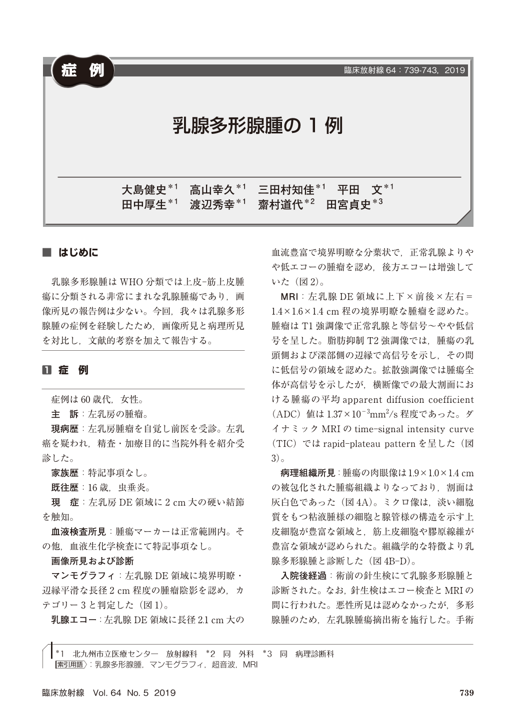
乳腺多形腺腫はWHO分類では上皮-筋上皮腫瘍に分類される非常にまれな乳腺腫瘍であり,画像所見の報告例は少ない。今回,我々は乳腺多形腺腫の症例を経験したため,画像所見と病理所見を対比し,文献的考察を加えて報告する。
Pleomorphic adenoma of the breast is a rare tumor, thus reports of image findings about this is rarely seen. Mammography showed a well-circumscribed lesion with smooth margin. Ultrasonography showed a well-circumscribed low-echoic mass. On magnetic resonance imaging(MRI), the tumor showed high signal intensity on T2-weighted images and rapid-plateau pattern on dynamic contrast-enhanced MRI. Histopathologic examination showed that the tumor was composed of an admixture of areas with abundant myxomatous cells and those with epithelial and myoepithelial components with fibrous stroma.
Copyright © 2019, KANEHARA SHUPPAN Co.LTD. All rights reserved.