Japanese
English
- 有料閲覧
PPV購入案内
1,650円 (1,500円+税10%) こちらは、206ページから209ページの文献です。購入には医書.jp本会員登録が必要です。
会員登録完了後に改めて上記「購入サイトへ」を選択してください。
または「購入サイトへ」選択後に「購入ログイン」、「新規会員登録」の順に進んでください。
会員登録について詳しくはをご確認ください。
- Abstract 文献概要
- 1ページ目 Look Inside
- 参考文献 Reference
Schloffer腫瘍は,手術瘢痕部の絹糸などの異物や肉芽腫を核として発生する遅発性・慢性炎症性結合組織性腫瘍である1).
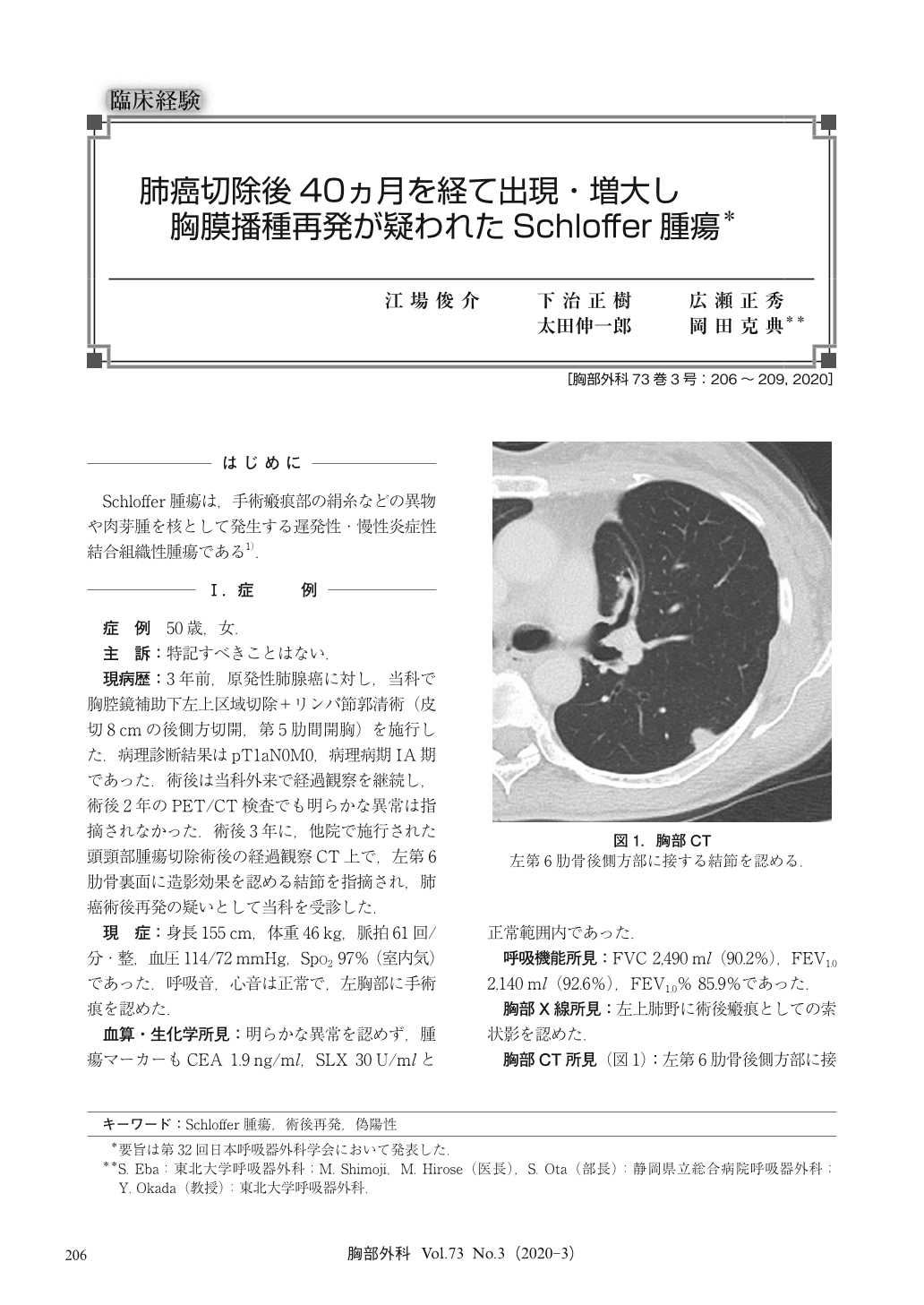
The case is a 50-year-old woman. Video-assisted thoracic surgery (VATS) left upper division segmentectomy was performed for adenocarcinoma through a postero-lateral incision. The pathological result was stage ⅠA. A chest computed tomography (CT) examination 40 months after the operation revealed a nodule in contact with the left 6th rib. A recurrence due to pleural dissemination was suspected in the PET/CT examination, but the possibility of reactive granulation was also considered. Two months later, chest CT reexamination showed an enlargement of the nodule, and reoperation was performed. A fibrous structure that appears to be a silk thread for closing chest at the 1st operation was found on the chest wall. The pathological examination revealed chronic suppurative pleuritis and organizing pneumonia without malignancy, leading to a diagnosis of Schloffer tumor.
© Nankodo Co., Ltd., 2020