Japanese
English
- 有料閲覧
PPV購入案内
1,650円 (1,500円+税10%) こちらは、574ページから577ページの文献です。購入には医書.jp本会員登録が必要です。
会員登録完了後に改めて上記「購入サイトへ」を選択してください。
または「購入サイトへ」選択後に「購入ログイン」、「新規会員登録」の順に進んでください。
会員登録について詳しくはをご確認ください。
- Abstract 文献概要
- 1ページ目 Look Inside
- 参考文献 Reference
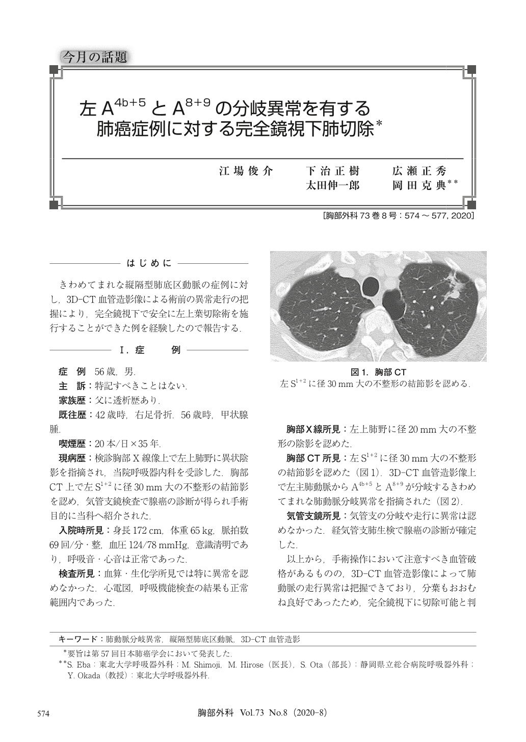
きわめてまれな縦隔型肺底区動脈の症例に対し,3D-CT血管造影像による術前の異常走行の把握により,完全鏡視下で安全に左上葉切除術を施行することができた例を経験したので報告する.
The case was a 56-year-old man. A nodular shadow of the left upper lobe was found in the chest computed tomography, and a diagnosis of adenocarcinoma was obtained by bronchoscopy. Preoperative 3-dimensional computed tomography (3D-CT) angiography indicated an extremely rare pulmonary artery bifurcation abnormality in which A4b+5 and A8+9 bifurcate from the left main pulmonary artery. Thoracoscopic left upper lobectomy and lymph node dissection were performed. Pathological diagnosis was adenocarcinoma with pStage ⅠB.
The mediastinal basal pulmonary artery is extremely rare, and to our knowledge, the bifurcation pattern of this case has not been reported elsewhere. The 3D-CT angiography was useful to detect the anatomical vascular abnormalities of the pulmonary artery before surgery, for the safe performance of the thoracoscopic surgery.
© Nankodo Co., Ltd., 2020