Japanese
English
- 有料閲覧
PPV購入案内
1,650円 (1,500円+税10%) こちらは、528ページから533ページの文献です。購入には医書.jp本会員登録が必要です。
会員登録完了後に改めて上記「購入サイトへ」を選択してください。
または「購入サイトへ」選択後に「購入ログイン」、「新規会員登録」の順に進んでください。
会員登録について詳しくはをご確認ください。
- Abstract 文献概要
- 1ページ目 Look Inside
- 参考文献 Reference
- サイト内被引用 Cited by
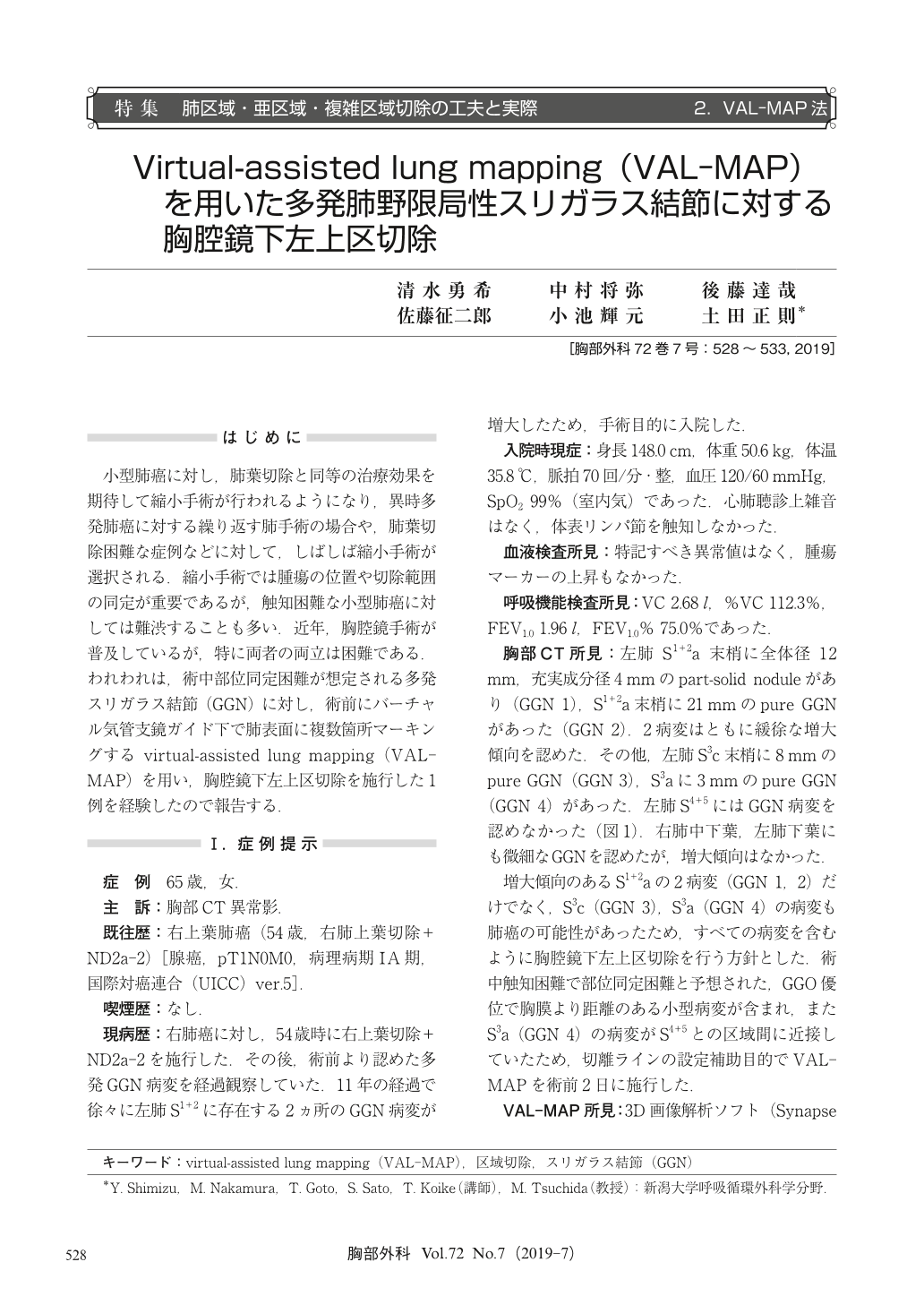
小型肺癌に対し,肺葉切除と同等の治療効果を期待して縮小手術が行われるようになり,異時多発肺癌に対する繰り返す肺手術の場合や,肺葉切除困難な症例などに対して,しばしば縮小手術が選択される.縮小手術では腫瘍の位置や切除範囲の同定が重要であるが,触知困難な小型肺癌に対しては難渋することも多い.近年,胸腔鏡手術が普及しているが,特に両者の両立は困難である.われわれは,術中部位同定困難が想定される多発スリガラス結節(GGN)に対し,術前にバーチャル気管支鏡ガイド下で肺表面に複数箇所マーキングするvirtual-assisted lung mapping(VAL-MAP)を用い,胸腔鏡下左上区切除を施行した1例を経験したので報告する.
Associated with an increase of small-sized lung cancer or metachronous second primary lung cancer, we have more opportunities to perform sublobar resection. Difficulties of identifying tumor location and appropriate surgical margin for small-sized ground-glass opacity (GGO) dominant lesions in thoracoscopic surgery is the big issue of sublobar resection. Virtual-assisted lung mapping (VAL-MAP) that makes markings on the lung surface through some peripheral bronchi by bronchoscopically projects intrapulmonary anatomy on the lung surface and literally draw a map. We report a case of thoracoscopic left upper division segmentectomy for multiple ground-glass nodules (GGNs) using preoperative VAL-MAP. A 65-year-old women who had undergone right upper lobectomy for primary lung cancer, and had multiple GGNs in the bilateral lungs was followed up as an outpatient. Eleven years after initial pulmonary resection, 2 lesions in the left upper division became bigger, and we decided to perform surgery for 4 GGNs in the left upper division including these 2 lesions. We preoperatively made bronchoscopic dye markings through B1+2c, B3a and B4a for in the left upper lobe. The 3 markings were intraoperatively identified. We decided the resection line based on the markings and performed thoracoscopic left upper division segmentectomy. The pathological diagnosis was minimally invasive adenocarcinoma, adenocarcinoma in situ and pneumonitis. Surgical margins were negative. VAL-MAP will assume an important role as an intraoperative navigation system for sublobar resection.
© Nankodo Co., Ltd., 2019