Japanese
English
- 有料閲覧
PPV購入案内
1,650円 (1,500円+税10%) こちらは、488ページから493ページの文献です。購入には医書.jp本会員登録が必要です。
会員登録完了後に改めて上記「購入サイトへ」を選択してください。
または「購入サイトへ」選択後に「購入ログイン」、「新規会員登録」の順に進んでください。
会員登録について詳しくはをご確認ください。
- Abstract 文献概要
- 1ページ目 Look Inside
- 参考文献 Reference
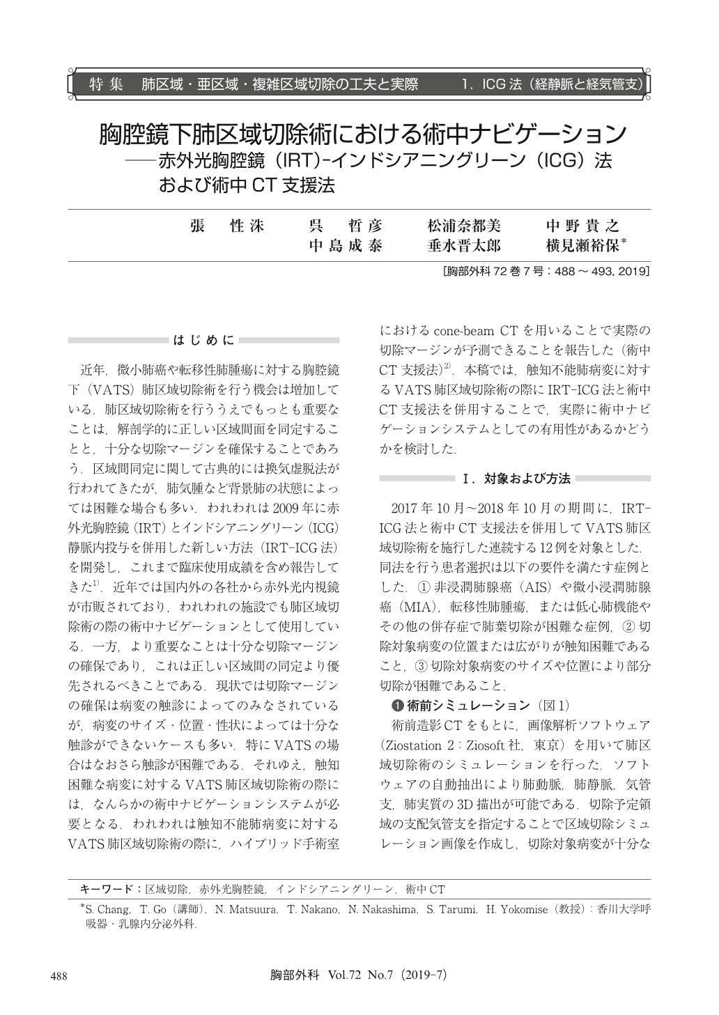
近年,微小肺癌や転移性肺腫瘍に対する胸腔鏡下(VATS)肺区域切除術を行う機会は増加している.肺区域切除術を行ううえでもっとも重要なことは,解剖学的に正しい区域間面を同定することと,十分な切除マージンを確保することであろう.区域間同定に関して古典的には換気虚脱法が行われてきたが,肺気腫など背景肺の状態によっては困難な場合も多い.われわれは2009年に赤外光胸腔鏡(IRT)とインドシアニングリーン(ICG)静脈内投与を併用した新しい方法(IRT-ICG法)を開発し,これまで臨床使用成績を含め報告してきた1).近年では国内外の各社から赤外光内視鏡が市販されており,われわれの施設でも肺区域切除術の際の術中ナビゲーションとして使用している.一方,より重要なことは十分な切除マージンの確保であり,これは正しい区域間の同定より優先されるべきことである.現状では切除マージンの確保は病変の触診によってのみなされているが,病変のサイズ・位置・性状によっては十分な触診ができないケースも多い.特にVATSの場合はなおさら触診が困難である.それゆえ,触知困難な病変に対するVATS肺区域切除術の際には,なんらかの術中ナビゲーションシステムが必要となる.われわれは触知不能肺病変に対するVATS肺区域切除術の際に,ハイブリッド手術室におけるcone-beam CTを用いることで実際の切除マージンが予測できることを報告した(術中CT支援法)2).本稿では,触知不能肺病変に対するVATS肺区域切除術の際にIRT-ICG法と術中CT支援法を併用することで,実際に術中ナビゲーションシステムとしての有用性があるかどうかを検討した.
Objectives:Recently, the use of video-assisted thoracoscopic surgery (VATS) segmentectomy for pulmonary malignancies has increased. For non-palpable lesions, securing a sufficient surgical margin is more likely to be uncertain. The purpose of this study was to evaluate the usefulness of our intraoperative navigation system in combination with the infrared thoracoscopy (IRT)-indocyanine green (ICG) method and intraoperative computed tomography (CT) during VATS segmentectomy for non-palpable pulmonary malignancies.
Methods:This study involved 12 consecutive patients who underwent both IRT-ICG and intraoperative CT-assisted thoracoscopic segmentectomy. Identification of the intersegmental line on the visceral pleura was visualized using IRT-ICG. The intersegmental line was marked by clipping, and intraoperative CT scan was performed under bilateral lung ventilation. The intraoperative CT images were used by the surgeons to confirm the correct anatomic segmental border and to secure a sufficient resection margin.
Results:A well-defined intersegmental line was observed in 83.3% of the patients. The rate of concordance between 3-dimensional (3D)-CT images reconstructed from intraoperative CT and preoperative simulation 3D-CT imaging was 91.7%. The mean surgical margin assessed on gross examination by the pathologist was 22.3 ± 4.5 mm. Complete resection was achieved in all patients using this approach.
Conclusions:Imaging support including preoperative simulation, IRT-ICG and intraoperative CT enables surgeons to perform definitive VATS segmentectomy for non-palpable lesions.
© Nankodo Co., Ltd., 2019