Japanese
English
- 有料閲覧
PPV購入案内
1,320円 (1,200円+税10%) こちらは、248ページから253ページの文献です。購入には医書.jp本会員登録が必要です。
会員登録完了後に改めて上記「購入サイトへ」を選択してください。
または「購入サイトへ」選択後に「購入ログイン」、「新規会員登録」の順に進んでください。
会員登録について詳しくはをご確認ください。
- Abstract 文献概要
- 1ページ目 Look Inside
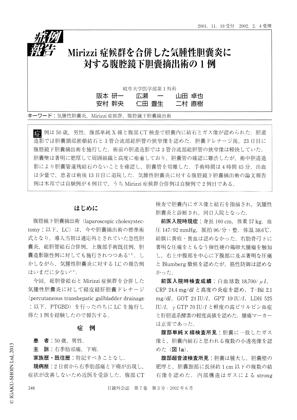
症例は50歳,男性.腹部単純X線と腹部CT検査で胆嚢内に結石とガス像が認められた.胆道造影では胆嚢頸部嵌頓結石と3管合流部総胆管の狭窄像を認めた.胆嚢ドレナージ後,23日目に腹腔鏡下胆嚢摘出術を施行した.術前の胆道造影では3管合流部総胆管の狭窄像は軽快していた.胆嚢壁は著明に肥厚して周囲組織と高度に癒着しており,胆嚢管の確認に難渋したが,術中胆道造影により胆嚢管遺残結石のないことを確認し,胆嚢管を切離した.手術時間は4時間45分,出血は少量で,患者は術後13日目に退院した.気腫性胆嚢炎に対する腹腔鏡下胆嚢例は本邦では自験例が6例目で,うちMirizzi症候群合併例は自験例で2例目である.
The patient was a 50-year-old man. Stones and the presence of gas in the gallbladder (GB) were detected by plain abdominal X-ray and computed tomography (CT) scan. Cholangiography showed an incarcerated stones in the cystic duct (CD) and a stenosis of the the common bile duct (CBD). Laparoscopic cholecystectomy (LC) was performed on the 23rd day after the percutaneous transhepatic gallbladder drainage. Pre-operative cholangiography showed improved stenosis of the CBD. The GB showed remarkable wall thickning and there was severely adhered to the peripheral tissues, making the CD hard to recognize.
Copyright © 2002, JAPAN SOCIETY FOR ENDOSCOPIC SURGERY All rights reserved.