Japanese
English
- 有料閲覧
PPV購入案内
1,320円 (1,200円+税10%) こちらは、691ページから694ページの文献です。購入には医書.jp本会員登録が必要です。
会員登録完了後に改めて上記「購入サイトへ」を選択してください。
または「購入サイトへ」選択後に「購入ログイン」、「新規会員登録」の順に進んでください。
会員登録について詳しくはをご確認ください。
- Abstract 文献概要
- 1ページ目 Look Inside
I.はじめに
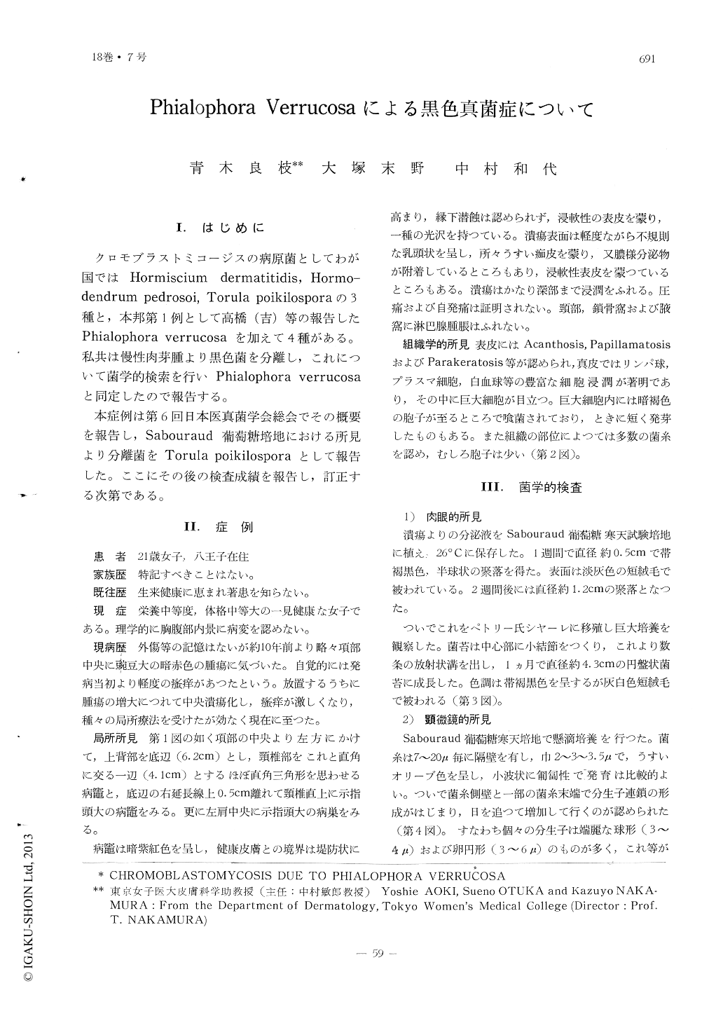
クロモブラストミコージスの病原菌としてわが国ではHormiscium dermatitidis, Hormo—dendrum pedrosoi, Torula poikilosporaの3種と,本邦第1例として高橋(吉)等の報告したPhialophora verrucosaを加えて4種がある。私共は慢性肉芽腫より黒色菌を分離し,これについて菌学的検索を行いPhialophora verrucosaと同定したので報告する。
本症例は第6回日本医真菌学会総会でその概要を報告し,Sabouraud葡萄糖培地における所見より分離菌をTorula poikilosporaとして報告した。ここにその後の検査成績を報告し,訂正する次第である。
Chromblastomycosis Due to Phialophora Verrucosa
Yoshie AOKI, Sueno OTUKA and Kazuyo NAKAMURA
From the Department of Dermatology,Tokyo Women's Medical College (Director :Prof. Dr. T. NAKAMURA)
Some ten years ago a 21-year-old female noted a lesion on the back of the neck, a which gradually enlarged and underwent ulceration.
On physical examination, a 6.2×4.1cm, raised, dull-red, indurated plaque on the back of the neck, showing a central ulceration with somewhat papillary vegetation was observed. In the area close to this lesion and on the left shoulder were two similar, finger tip-sized lesions.
Histological examination revealed in the cutis a granulomatous infiltrate, composed of leucocytes, epithelioid cells, giant cells, lym-phocytes and plasma cells. The giant cells contained round, thick-walled, dark brown spores, some of which showed a short budding, In some areas of the affected tissue there were found not only spores but abundant hyphae.
Cultures on Sabouraucl's glucose agar yielded colonies which were dark brown to black in color with a grey short aerial mycelium.Microscopically, on Sabouraud's glucose agar oval to spherical conidia were borne in long branching chain formation mainly from the sides of the hyphae. Conidia in some of the chains were so irregular in shape that the conidial chains resembled those of Torula poikilospora. On corn meal glucose agar co-nidia were produced from cups at the tip of flask-shaped conidiophores.
Mice injected intraperitoneally with a sus-pension of culture showed numerous black granulomatous nodules scattered throughout the peritoneum.
The organism has been identified as Phialo-phora verrucosa.
Copyright © 1964, Igaku-Shoin Ltd. All rights reserved.