Japanese
English
- 有料閲覧
PPV購入案内
1,320円 (1,200円+税10%) こちらは、423ページから427ページの文献です。購入には医書.jp本会員登録が必要です。
会員登録完了後に改めて上記「購入サイトへ」を選択してください。
または「購入サイトへ」選択後に「購入ログイン」、「新規会員登録」の順に進んでください。
会員登録について詳しくはをご確認ください。
- Abstract 文献概要
- 1ページ目 Look Inside
I.はじめに
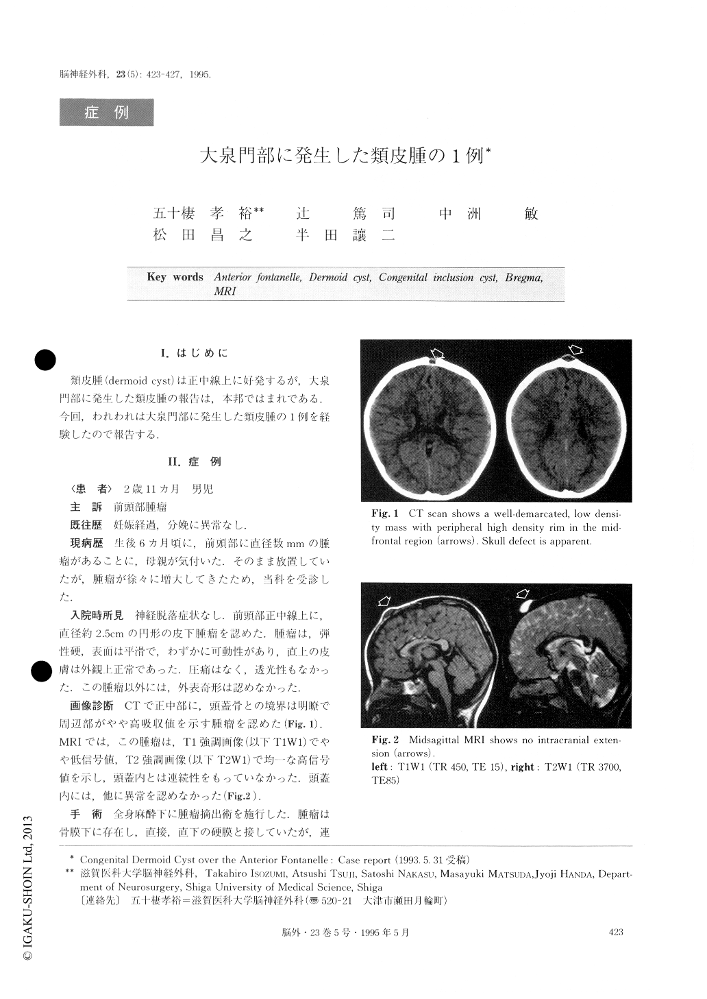
類皮腫(dermoid cyst)は正中線上に好発するが,大泉門部に発生した類皮腫の報告は,本邦ではまれである.今回,われわれは大泉門部に発生した類皮腫の1例を経験したので報告する.
We report a case of a 35-month-old Japanese boy harboring a dermoid cyst in the region of the anterior fontanelle, which is rarely reported among Japanese. A small soft subcutaneous mass was noticed in the mid-frontal region at the age of 6 months. On admission, asoft, nontender round mass (2.5cm in diameter) was lo-cated over the anterior fontanelle. Transillumination was negative. Neurological examination yielded normal findings. MRI showed a well-circumscribed mass, which was slightly hypointense on T1W1 and hyperin-tense on T2W1. Midsagittal MRI showed no intracra-nial extension.
At operation, a well-encapsulated mass was found beneath the pericranium, which was easily dissected from the underlying dura mater and removed. The tumor contained caseous material and several hairs. Microscopically, the cyst wall consisted of connective tissues lined by stratified squamous epithelium. Sebaceous glands and hair follicles were found in the subepithelial layer.
It is important to confirm preoperatively whether or not the extracranial mass extends into the intracranial cavity. For this purpose, midsagittal or coronal MRI seems to be most useful.
Copyright © 1995, Igaku-Shoin Ltd. All rights reserved.