Japanese
English
- 有料閲覧
PPV購入案内
1,320円 (1,200円+税10%) こちらは、789ページから793ページの文献です。購入には医書.jp本会員登録が必要です。
会員登録完了後に改めて上記「購入サイトへ」を選択してください。
または「購入サイトへ」選択後に「購入ログイン」、「新規会員登録」の順に進んでください。
会員登録について詳しくはをご確認ください。
- Abstract 文献概要
- 1ページ目 Look Inside
I.はじめに
最近,わが国においても脳腫瘍や脳動静脈奇形に対してradiosurgeryが数多く実施されている.脳腫瘍に対しては当初は主に良性腫瘍が治療の対象となっていたが,転移性脳腫瘍に対しての報告2-7)もしだいに増加し,その治療効果も認められつつある.
しかし,radiosurgeryの合併症については,治療後の脳浮腫の増強による神経症状の増悪や遅発性の放射線壊死による新たな神経症状の出現等が報告2,3)されているが,radiosurgeryに関連した腫瘍出血の報告は見当たらない.
An unusual case of peritumoral hemorrhage after radiosurgery for the treatment of metastatic brain tumor is reported. This 64-year-old woman had a his-tory of breast cancer and underwent right mastectomy in 1989. She remained well until January 1993, when she started to have headache, nausea and speech dis-turbance, and was hospitalized on February 25, 1993. Neurological examination disclosed right hemiparesis and bilateral papilledema.
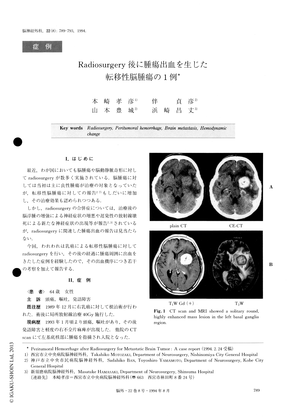
CT scan and MR imaging showed a solitary round mass lesion in the left basal ganglia region. It was a well-demarcated, highly enhanced mass, 37mm in dia-meter. Cerebral angiography confirmed a highly vascular mass lesion in the same location.
She was treated with radiosurgery on March 8 (maxium dose was 20Gy in the center and 10Gy in the peripheral part of the tumor). After radiosurgery, she had an uneventful course and clinical and radiosurgical improvement could be detected. Her neurological symp-toms and signs gradually improved and reduction of the tumor size and perifocal edema could be seen one month after radiosurgery. However, 6 weeks after radiosurgery, she suddenly developed semicoma and right hemiplegia. CT scan disclosed a massive peritu-moral hemorrhage. Then, emergency craniotomy, eva-cuation of the hematoma and total removal of the tumor were performed on April 24. Histopathological diagnosis was adenocarcinoma. It was the same finding as that of the previous breast cancer. Histopathological examination revealed necrosis without tumor cells in the center and residual tumor cells in the peripheral part of the tumor.
It is postulated that peritumoral hemorrhage was caused by hemodynamic changes in the vascular-rich tumor after radiosurgery and breakdown of the fragile abnormal vessels in the peripheral part of the tumor.
Copyright © 1994, Igaku-Shoin Ltd. All rights reserved.