Japanese
English
- 有料閲覧
- Abstract 文献概要
- 1ページ目 Look Inside
I.はじめに
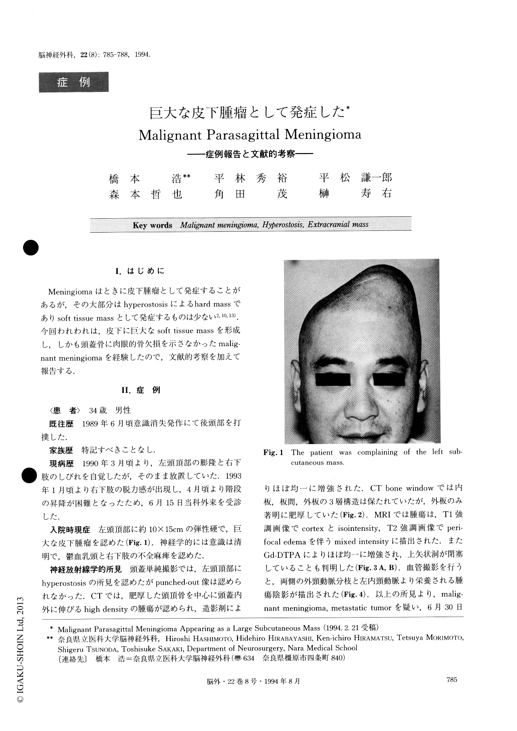
Meningiomaはときに皮下腫瘤として発症することがあるが,その大部分はhyperostosisによるhard massでありsoft tissue massとして発症するものは少ない2,10,13).今回われわれは,皮下に巨大なsoft tissue massを形成し,しかも頭蓋骨に肉眼的骨欠損を示さなかったmalg—nant meningiomaを経験したので,文献的考察を加えて報告する.
We present a rare case of malignant meningioma appearing as an extracranial soft-tissue mass.
A 34-year-old male was admitted with left parietal subcutaneous soft-tissue mass. Neurological examina-tion on admission revealed right leg monoparesis and papilledema. Skull X rays showed hyperostosis of the left parietal bone. There was no evidence of bony des-truction. CT and MRI showed a tumor with a large in-tracranial component, which was homogeneously en-hanced. The tumor was totally removed by left osto-plastic craniotomy. The light-microscopic examination showed a meningothelial meningioma accompanied by malignant components with necrotic foci and mitotic fi-gures. Tumor cells had invaded all the layers of the skull via the Haversian canals. We propose that hyper- ostosis with tumor invasion through all three layers of the skull should be considered as a malignant feature.

Copyright © 1994, Igaku-Shoin Ltd. All rights reserved.


