Japanese
English
- 有料閲覧
PPV購入案内
1,320円 (1,200円+税10%) こちらは、677ページから680ページの文献です。購入には医書.jp本会員登録が必要です。
会員登録完了後に改めて上記「購入サイトへ」を選択してください。
または「購入サイトへ」選択後に「購入ログイン」、「新規会員登録」の順に進んでください。
会員登録について詳しくはをご確認ください。
- Abstract 文献概要
- 1ページ目 Look Inside
I.はじめに
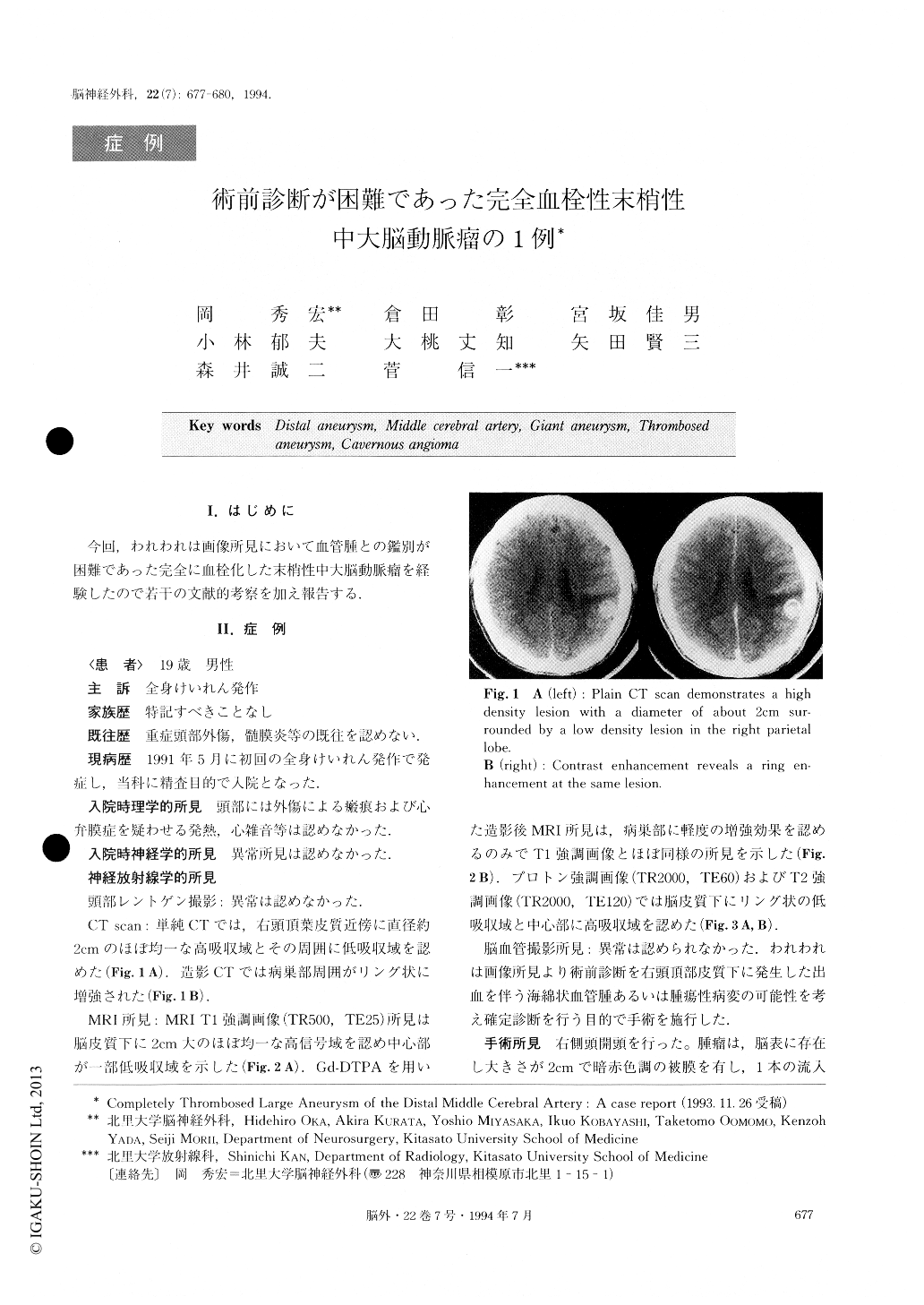
今回,われわれは画像所見において血管腫との鑑別が困難であった完全に血栓化した末梢性中大脳動脈瘤を経験したので若干の文献的考察を加え報告する.
A 19 year old male was admitted for evaluation after a seizure. Physical and neurological examination was normal. CT demonstrated an en larged, high density mass in the right parietal lobe. MRI showed a homogeneous high intensity T1 weighted mass, sur-rounded by a low intensity T2 weighted rim in the right parietal lobe. Angiography did not show any abnormal findings. A diagnosis of cavernous angioma with primary bleeding in the subcortical region of the right parietal lobe was made after radiological examina-tion.
Histological examination showed a completely throm-hosed aneurysm. The mechanism of the complete thrombosis and the growth of this large aneurysm and the shortcomings of radiological examination are discussed.
Copyright © 1994, Igaku-Shoin Ltd. All rights reserved.