Japanese
English
- 有料閲覧
PPV購入案内
1,320円 (1,200円+税10%) こちらは、285ページから289ページの文献です。購入には医書.jp本会員登録が必要です。
会員登録完了後に改めて上記「購入サイトへ」を選択してください。
または「購入サイトへ」選択後に「購入ログイン」、「新規会員登録」の順に進んでください。
会員登録について詳しくはをご確認ください。
- Abstract 文献概要
- 1ページ目 Look Inside
I.はじめに
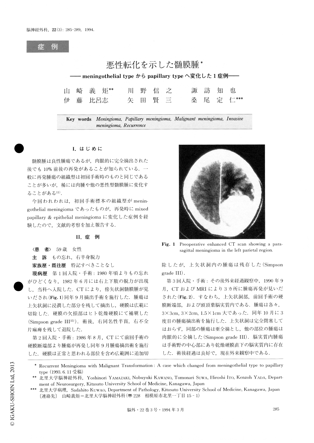
髄膜腫は良性腫瘍であるが,肉眼的に完全摘出された後でも10%前後の再発があることが知られている.一般に再発腫瘍の組織型は初回手術時のものと同じであることが多いが,稀には肉腫や他の悪性型髄膜腫に変化することがある11).
今回われわれは,初回手術標本の組織型がmenin—gothelial meningiomaであったものが,再発時にmixed papillary & epithelial meningiomaに変化した症例を経験したので,文献的考察を加え報告する.
We report here a case with meningioma showing malignant transformation in its course of multiple re-currences. A 59-year-old woman developed a right-sided hemiparesis in June, 1982 and CT scan disclosed a parasagittal well-enhanced mass. The tumor was sub-totally removed (Simpson grade Ⅲ) by an operation in September, 1982. Histological findings of the tumor were consistent with a meningothelial meningioma but showed no malignant features, such as high cellularity, necrotic foci, high mitotic rate, or nuclear pleomor-phism. However, the tumor did invade the underlying cerebral cortex. In August, 1986, a recurrent tumor was detected by CT scan and was removed (Simpson grade Ⅲ). The tumor tissue at the second operation showed the same histological features as the first specimen. In September, 1990, the patient developed multiple in-tracranial recurrences. There were three tumor nodules, all of which were removed. Histologically, significant histological differences between the second and the third operative specimens were found. In the last tumor tissue, one nodule showed a papillary pattern. In the other tumor nodules, each tumor cell had proliferated separately instead of adhering to other tumor cells to form a syncitium. This histological pattern was consis-tent with an epithelial meningioma described by Cushing and Eisenhardt in 1938. The papillary portion of the tumor was stained with monoclonal antibody Ki-67 in frozen section. The labelling index was 9.7%, which was as high as malignant meningioma. Electron microscopic examination of the papillary portion of the tumor showed that the tumor cells had irregular nuclei, interdigitations between the adjacent plasma mem-branes and a few ill-developed desmosomes. These findings supported the suspicion of the malignant char-acter of the papillary meningioma.
Copyright © 1994, Igaku-Shoin Ltd. All rights reserved.