Japanese
English
- 有料閲覧
PPV購入案内
1,320円 (1,200円+税10%) こちらは、579ページから586ページの文献です。購入には医書.jp本会員登録が必要です。
会員登録完了後に改めて上記「購入サイトへ」を選択してください。
または「購入サイトへ」選択後に「購入ログイン」、「新規会員登録」の順に進んでください。
会員登録について詳しくはをご確認ください。
- Abstract 文献概要
- 1ページ目 Look Inside
- 参考文献 Reference
Point
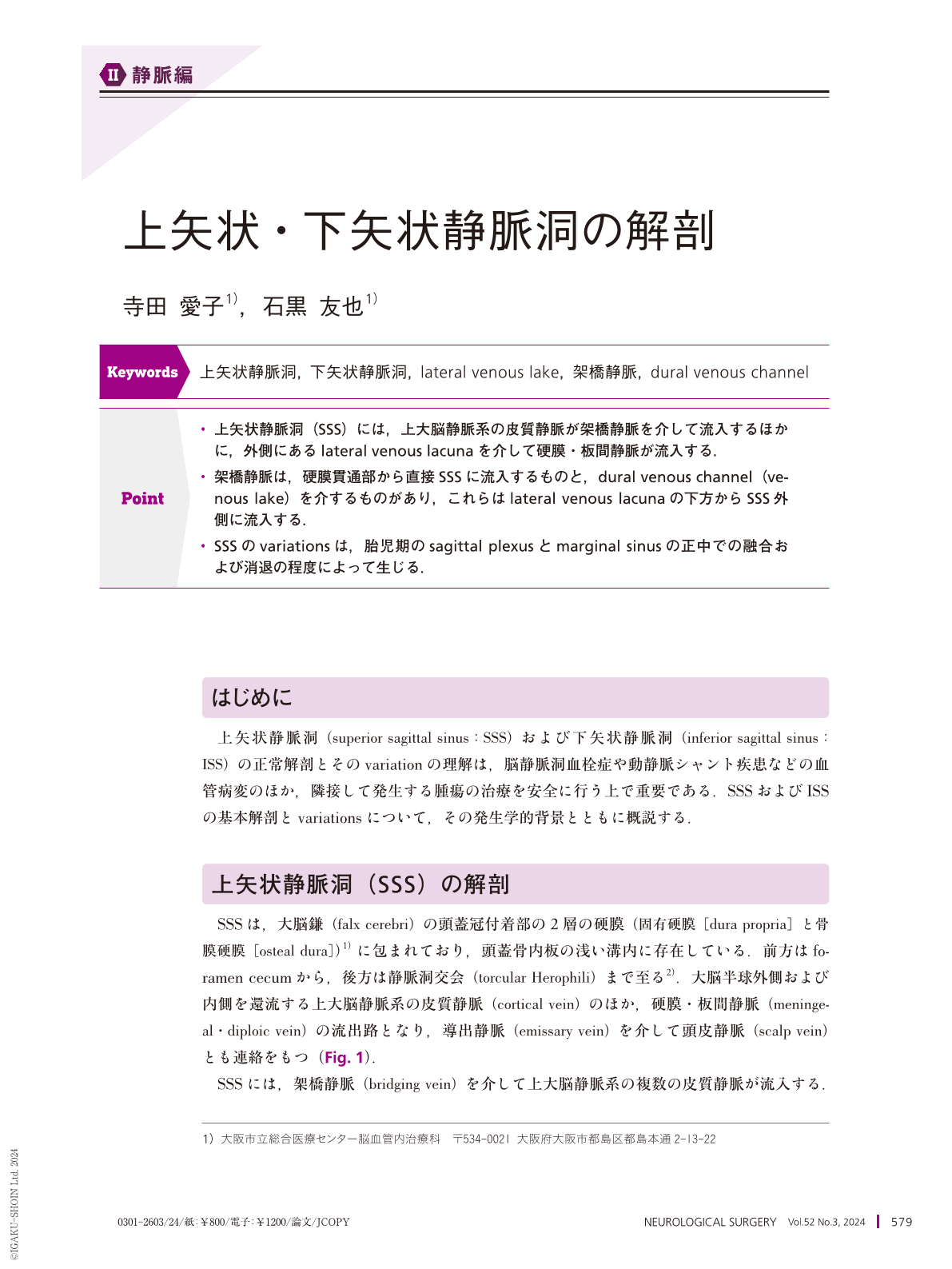
・上矢状静脈洞(SSS)には,上大脳静脈系の皮質静脈が架橋静脈を介して流入するほかに,外側にあるlateral venous lacunaを介して硬膜・板間静脈が流入する.
・架橋静脈は,硬膜貫通部から直接SSSに流入するものと,dural venous channel(venous lake)を介するものがあり,これらはlateral venous lacunaの下方からSSS外側に流入する.
・SSSのvariationsは,胎児期のsagittal plexusとmarginal sinusの正中での融合および消退の程度によって生じる.
The superior sagittal sinus(SSS)is contained within the dura, which consists of the dura propria and osteal dura at the junction of the falx cerebri, in addition to the attachment of the falx to the cranial vault. The SSS extends anteriorly from the foramen cecum and posteriorly to the torcular Herophili. The superior cerebral veins flow into the SSS, coursing under the lateral venous lacunae via bridging veins. Most of the bridging veins reach the dura and empty directly into the SSS. However, some are attached to the dural or existed in it for some distance before their sinus entrance. The venous structures of the junctional zone between the bridging vein and the SSS existed in the dura are referred to as dural venous channels. The SSS communicates with the lateral venous lacunae connecting the meningeal and diploic veins, as well as the emissary veins. These anatomical variations of the SSS are defined by the embryological processes of fusion and withdrawal of the sagittal plexus and marginal sinus.
Copyright © 2024, Igaku-Shoin Ltd. All rights reserved.