Japanese
English
- 有料閲覧
PPV購入案内
1,320円 (1,200円+税10%) こちらは、859ページから867ページの文献です。購入には医書.jp本会員登録が必要です。
会員登録完了後に改めて上記「購入サイトへ」を選択してください。
または「購入サイトへ」選択後に「購入ログイン」、「新規会員登録」の順に進んでください。
会員登録について詳しくはをご確認ください。
- Abstract 文献概要
- 1ページ目 Look Inside
- 参考文献 Reference
Ⅰ.はじめに
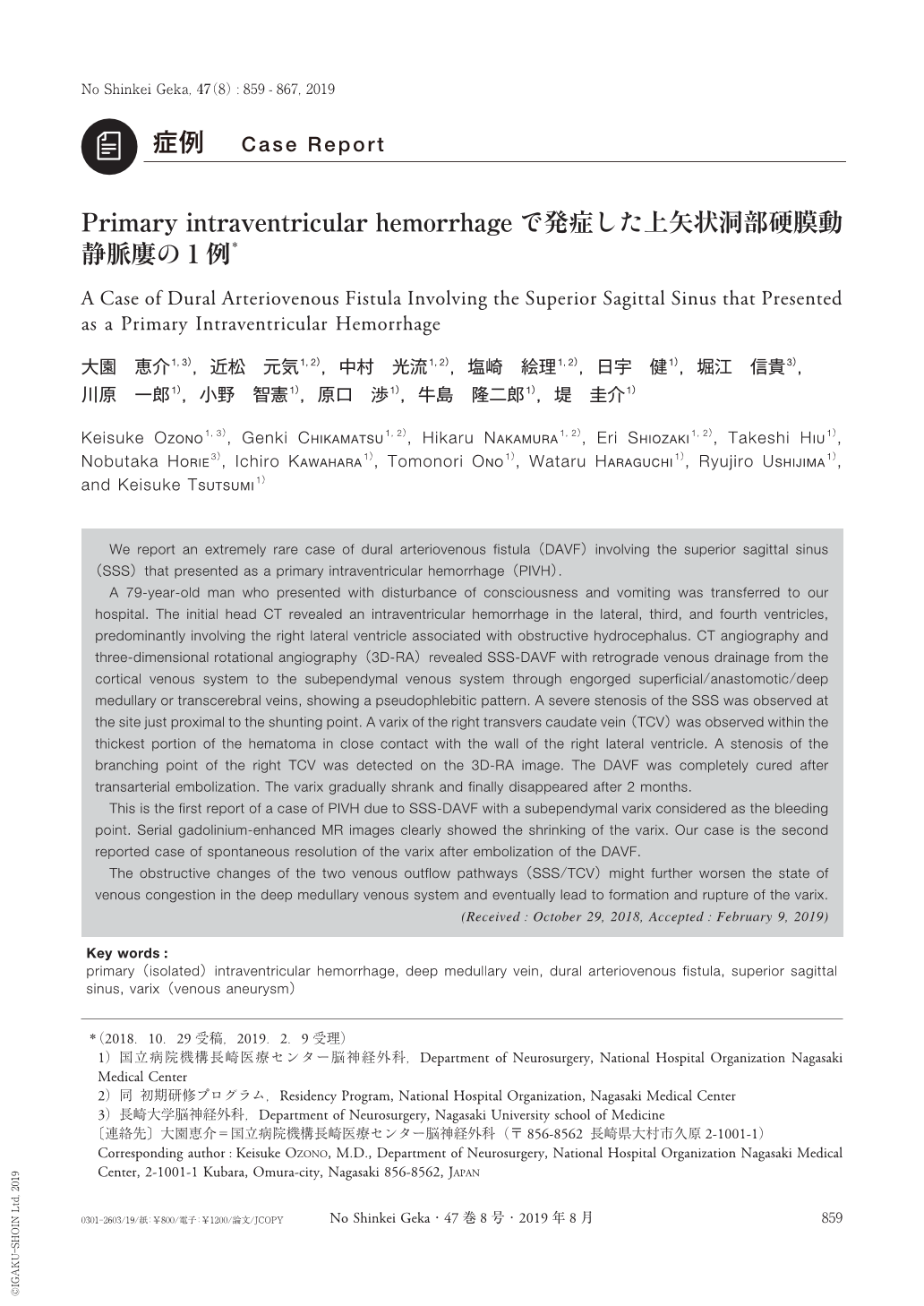
硬膜動静脈廔(dural arteriovenous fistula:DAVF)が純粋な脳室内出血(primary intraventricular hemorrhage:PIVH)で発症することは稀であり3,7),報告例の中では横-S状静脈洞部(transvers-sigmoid sinus:TSS)DAVFが多くを占める2).今回われわれは,PIVHで発症した上矢状洞部(superior sagittal sinus:SSS)DAVFの極めて稀な1例を経験したので,文献的考察を加えて報告する.
We report an extremely rare case of dural arteriovenous fistula(DAVF)involving the superior sagittal sinus(SSS)that presented as a primary intraventricular hemorrhage(PIVH).
A 79-year-old man who presented with disturbance of consciousness and vomiting was transferred to our hospital. The initial head CT revealed an intraventricular hemorrhage in the lateral, third, and fourth ventricles, predominantly involving the right lateral ventricle associated with obstructive hydrocephalus. CT angiography and three-dimensional rotational angiography(3D-RA)revealed SSS-DAVF with retrograde venous drainage from the cortical venous system to the subependymal venous system through engorged superficial/anastomotic/deep medullary or transcerebral veins, showing a pseudophlebitic pattern. A severe stenosis of the SSS was observed at the site just proximal to the shunting point. A varix of the right transvers caudate vein(TCV)was observed within the thickest portion of the hematoma in close contact with the wall of the right lateral ventricle. A stenosis of the branching point of the right TCV was detected on the 3D-RA image. The DAVF was completely cured after transarterial embolization. The varix gradually shrank and finally disappeared after 2 months.
This is the first report of a case of PIVH due to SSS-DAVF with a subependymal varix considered as the bleeding point. Serial gadolinium-enhanced MR images clearly showed the shrinking of the varix. Our case is the second reported case of spontaneous resolution of the varix after embolization of the DAVF.
The obstructive changes of the two venous outflow pathways(SSS/TCV)might further worsen the state of venous congestion in the deep medullary venous system and eventually lead to formation and rupture of the varix.
Copyright © 2019, Igaku-Shoin Ltd. All rights reserved.