Japanese
English
- 有料閲覧
PPV購入案内
1,320円 (1,200円+税10%) こちらは、508ページから515ページの文献です。購入には医書.jp本会員登録が必要です。
会員登録完了後に改めて上記「購入サイトへ」を選択してください。
または「購入サイトへ」選択後に「購入ログイン」、「新規会員登録」の順に進んでください。
会員登録について詳しくはをご確認ください。
- Abstract 文献概要
- 1ページ目 Look Inside
- 参考文献 Reference
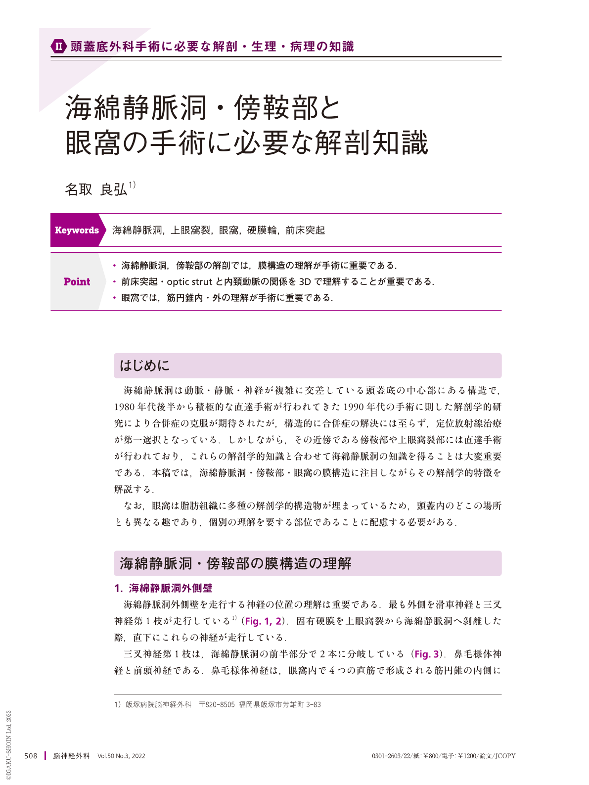
Point
・海綿静脈洞,傍鞍部の解剖では,膜構造の理解が手術に重要である.
・前床突起・optic strutと内頚動脈の関係を3Dで理解することが重要である.
・眼窩では,筋円錐内・外の理解が手術に重要である.
The cavernous sinus, para-sellar region, and orbit have intricately intertwined cranial nerves, blood vessels, and dura mater. In surgery, anatomical understanding is very important. Recognizing the location(depth)of the cranial nerves running on the lateral and upper wall of the cavernous sinus is vital and is directly linked to postoperative complications. In addition, understanding the dural ring in the clinoid segment of the internal carotid artery is important. The periosteum on the upper surface of the anterior clinoid is the distal dural ring of the internal carotid artery, and the periosteum on the lower surface is the proximal dural ring. The orbit is filled with adipose tissue and is completely different from other intracranial parts. However, understanding the anatomy from the orbital apex to the superior orbital fissure is important in the pterional approach.
Copyright © 2022, Igaku-Shoin Ltd. All rights reserved.