Japanese
English
- 有料閲覧
PPV購入案内
1,320円 (1,200円+税10%) こちらは、16ページから23ページの文献です。購入には医書.jp本会員登録が必要です。
会員登録完了後に改めて上記「購入サイトへ」を選択してください。
または「購入サイトへ」選択後に「購入ログイン」、「新規会員登録」の順に進んでください。
会員登録について詳しくはをご確認ください。
- Abstract 文献概要
- 1ページ目 Look Inside
- 参考文献 Reference
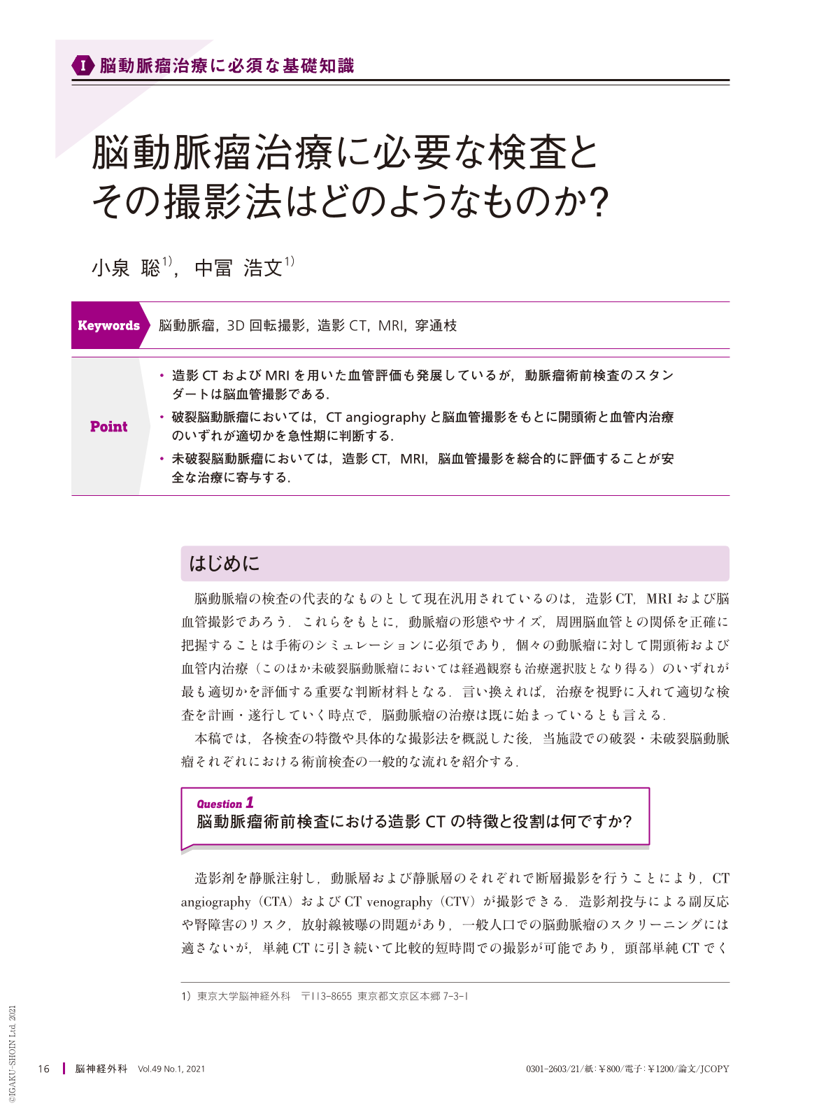
Point
・造影CTおよびMRIを用いた血管評価も発展しているが,動脈瘤術前検査のスタンダートは脳血管撮影である.
・破裂脳動脈瘤においては,CT angiographyと脳血管撮影をもとに開頭術と血管内治療のいずれが適切かを急性期に判断する.
・未破裂脳動脈瘤においては,造影CT,MRI,脳血管撮影を総合的に評価することが安全な治療に寄与する.
Acquiring appropriate preoperative images is an important step in the treatment of cerebral aneurysms. Despite recent advances in contrast-enhanced CT and MRI, catheter angiography remains the standard of care in preoperative imaging tests for both ruptured and unruptured intracranial aneurysms. Three-dimensional rotational angiography can provide a clear view of vascular structure around the aneurysm in an intuitive manner, including the small perforators. For ruptured aneurysms, the treatment modality(i.e., surgical clipping or endovascular embolization)is usually based on emergent contrast CT and catheter angiography findings. For unruptured aneurysms, integrated assessment involving CT, MRI, and angiography is often useful in multimodal treatment decision making.
Copyright © 2021, Igaku-Shoin Ltd. All rights reserved.