Japanese
English
- 有料閲覧
PPV購入案内
1,320円 (1,200円+税10%) こちらは、555ページから560ページの文献です。購入には医書.jp本会員登録が必要です。
会員登録完了後に改めて上記「購入サイトへ」を選択してください。
または「購入サイトへ」選択後に「購入ログイン」、「新規会員登録」の順に進んでください。
会員登録について詳しくはをご確認ください。
- Abstract 文献概要
- 1ページ目 Look Inside
- 参考文献 Reference
Ⅰ.はじめに
2014年1月,本邦で脳神経領域の動脈塞栓術においてエンボスフィア®(日本化薬,東京)の使用が保険適用となった.ゼラチンでコーティングされたアクリル系共重合体からなる非吸水性マイクロスフィアで,今後塞栓物質の中心的な役割を担うと考えられている.当施設では巨大な髄膜腫の摘出術前に塞栓術を実施しており,今回,エンボスフィア®を用いた塞栓術後の初期成績について検討したため報告する.
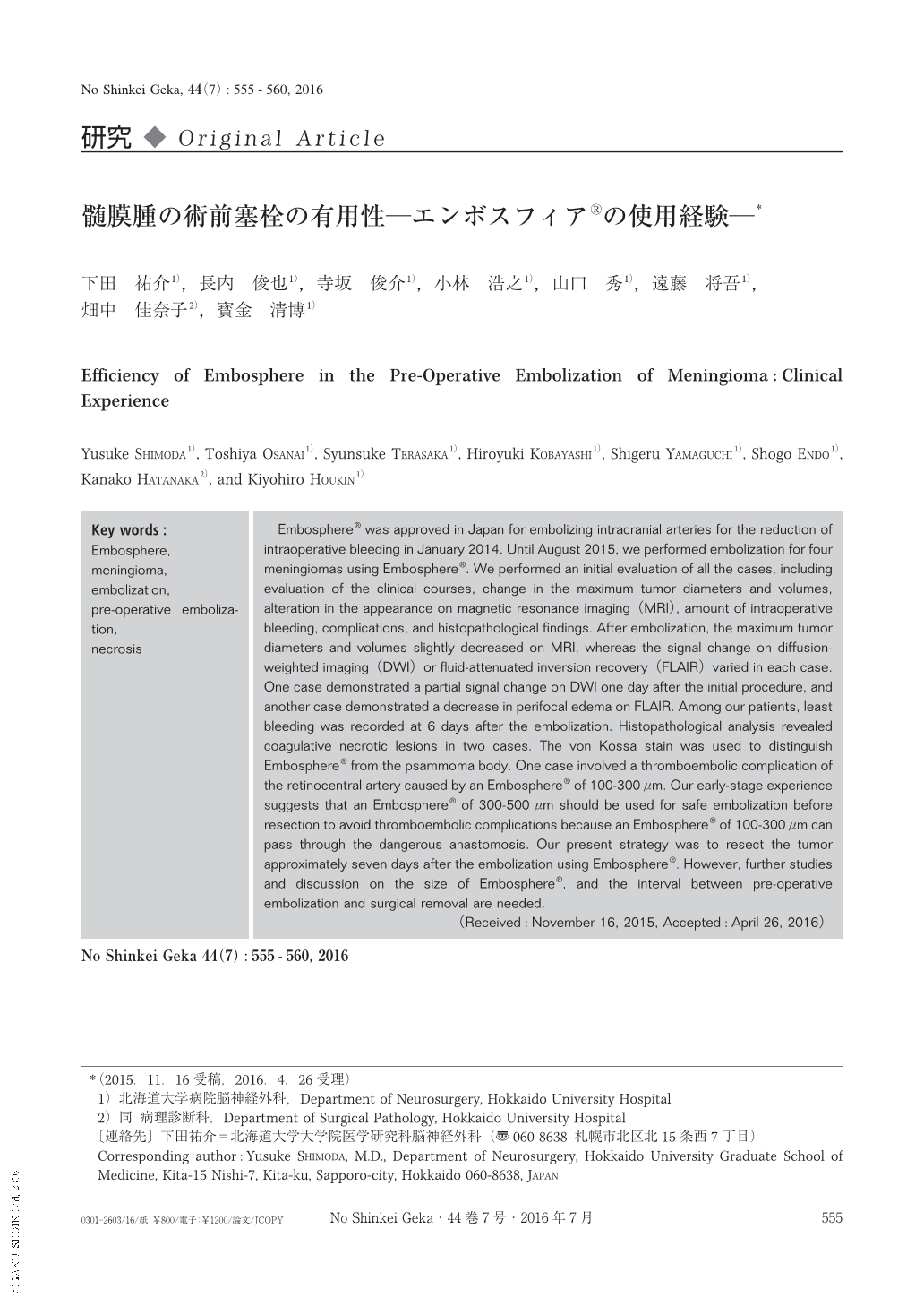
Embosphere® was approved in Japan for embolizing intracranial arteries for the reduction of intraoperative bleeding in January 2014. Until August 2015, we performed embolization for four meningiomas using Embosphere®. We performed an initial evaluation of all the cases, including evaluation of the clinical courses, change in the maximum tumor diameters and volumes, alteration in the appearance on magnetic resonance imaging(MRI), amount of intraoperative bleeding, complications, and histopathological findings. After embolization, the maximum tumor diameters and volumes slightly decreased on MRI, whereas the signal change on diffusion-weighted imaging(DWI)or fluid-attenuated inversion recovery(FLAIR)varied in each case. One case demonstrated a partial signal change on DWI one day after the initial procedure, and another case demonstrated a decrease in perifocal edema on FLAIR. Among our patients, least bleeding was recorded at 6 days after the embolization. Histopathological analysis revealed coagulative necrotic lesions in two cases. The von Kossa stain was used to distinguish Embosphere® from the psammoma body. One case involved a thromboembolic complication of the retinocentral artery caused by an Embosphere® of 100-300 μm. Our early-stage experience suggests that an Embosphere® of 300-500 μm should be used for safe embolization before resection to avoid thromboembolic complications because an Embosphere® of 100-300 μm can pass through the dangerous anastomosis. Our present strategy was to resect the tumor approximately seven days after the embolization using Embosphere®. However, further studies and discussion on the size of Embosphere®, and the interval between pre-operative embolization and surgical removal are needed.
Copyright © 2016, Igaku-Shoin Ltd. All rights reserved.