Japanese
English
- 有料閲覧
- Abstract 文献概要
- 1ページ目 Look Inside
- 参考文献 Reference
Ⅰ.はじめに
グロームス腫瘍は全身の傍神経節から発生する良性腫瘍である.頸静脈孔部グロームス腫瘍は脳神経外科医が治療する代表的部位であるが,手術治療には下位脳神経を含めた複数の脳神経障害や頸動脈損傷を惹起する可能性がある.さまざまな頭蓋底到達法を用いたとしても全摘出は必ずしも容易ではない1,2,7,14,19,23).またグロームス腫瘍には,数%にカテコラミン分泌を認め,褐色細胞腫と同様の臨床症状を呈するものがある.これらに外科治療を行う場合には循環血漿量の減少状態や術中のカテコラミン過剰分泌は大きな問題で,術後の低血圧や低血糖にも適切に対処しなければならない9, 21).
今回われわれは,カテコラミン分泌型頸静脈孔部グロームス腫瘍の1例を経験し,良好な治療経過を得た.本腫瘍は頸静脈孔部グロームス腫瘍とカテコラミン分泌腫瘍の両方の特徴を有しており適切な治療戦略と周術期管理が必要である.文献的考察を加えわれわれの本腫瘍に対する治療戦略と周術期管理を報告する.
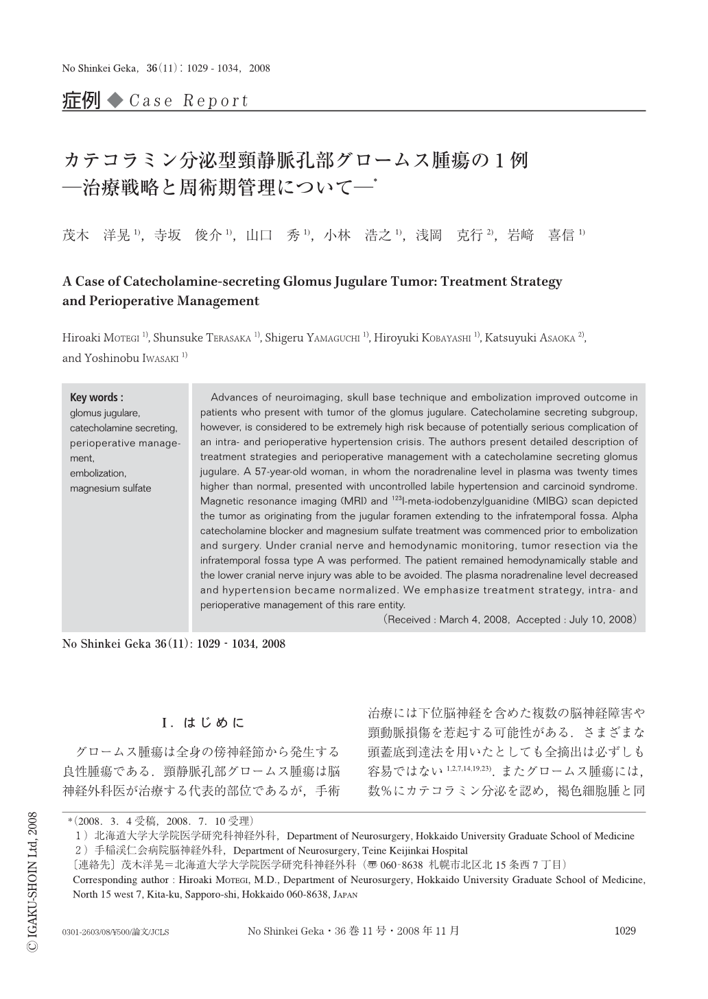
Advances of neuroimaging, skull base technique and embolization improved outcome in patients who present with tumor of the glomus jugulare. Catecholamine secreting subgroup, however, is considered to be extremely high risk because of potentially serious complication of an intra- and perioperative hypertension crisis. The authors present detailed description of treatment strategies and perioperative management with a catecholamine secreting glomus jugulare. A 57-year-old woman, in whom the noradrenaline level in plasma was twenty times higher than normal, presented with uncontrolled labile hypertension and carcinoid syndrome. Magnetic resonance imaging (MRI) and 123I-meta-iodobenzylguanidine (MIBG) scan depicted the tumor as originating from the jugular foramen extending to the infratemporal fossa. Alpha catecholamine blocker and magnesium sulfate treatment was commenced prior to embolization and surgery. Under cranial nerve and hemodynamic monitoring, tumor resection via the infratemporal fossa type A was performed. The patient remained hemodynamically stable and the lower cranial nerve injury was able to be avoided. The plasma noradrenaline level decreased and hypertension became normalized. We emphasize treatment strategy, intra- and perioperative management of this rare entity.

Copyright © 2008, Igaku-Shoin Ltd. All rights reserved.


