Japanese
English
- 有料閲覧
PPV購入案内
1,320円 (1,200円+税10%) こちらは、731ページから735ページの文献です。購入には医書.jp本会員登録が必要です。
会員登録完了後に改めて上記「購入サイトへ」を選択してください。
または「購入サイトへ」選択後に「購入ログイン」、「新規会員登録」の順に進んでください。
会員登録について詳しくはをご確認ください。
- Abstract 文献概要
- 1ページ目 Look Inside
- 参考文献 Reference
Ⅰ.はじめに
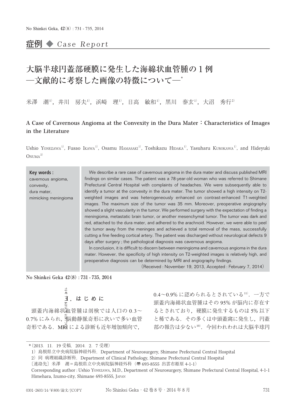
頭蓋内海綿状血管腫は剖検では人口の0.3~0.7%にみられ,脳動静脈奇形に次いで多い血管奇形である.MRIによる診断も近年増加傾向で,0.4~0.9%に認められるとされている11).一方で頭蓋内海綿状血管腫はその95%が脳内に存在するとされており,硬膜に発生するものは5%以下と稀である.その多くは中頭蓋窩に発生し,円蓋部の報告は少ない10).今回われわれは大脳半球円蓋部硬膜に発生した海綿状血管腫の1例を経験したので画像上の特徴について文献的考察を加えて報告する.
We describe a rare case of cavernous angioma in the dura mater and discuss published MRI findings on similar cases. The patient was a 78-year-old woman who was referred to Shimane Prefectural Central Hospital with complaints of headaches. We were subsequently able to identify a tumor at the convexity in the dura mater. The tumor showed a high intensity on T2-weighted images and was heterogeneously enhanced on contrast-enhanced T1-weighted images. The maximum size of the tumor was 35 mm. Moreover, preoperative angiography showed a slight vascularity in the tumor. We performed surgery with the expectation of finding a meningioma, metastatic brain tumor, or another mesenchymal tumor. The tumor was dark and red, attached to the dura mater, and adhered to the arachnoid. However, we were able to peel the tumor away from the meninges and achieved a total removal of the mass, successfully cutting a fine feeding cortical artery. The patient was discharged without neurological defects 9 days after surgery;the pathological diagnosis was cavernous angioma.
In conclusion, it is difficult to discern between meningioma and cavernous angioma in the dura mater. However, the specificity of high intensity on T2-weighted images is relatively high, and preoperative diagnosis can be determined by MRI and angiography findings.
Copyright © 2014, Igaku-Shoin Ltd. All rights reserved.