Japanese
English
- 有料閲覧
PPV購入案内
1,320円 (1,200円+税10%) こちらは、297ページから300ページの文献です。購入には医書.jp本会員登録が必要です。
会員登録完了後に改めて上記「購入サイトへ」を選択してください。
または「購入サイトへ」選択後に「購入ログイン」、「新規会員登録」の順に進んでください。
会員登録について詳しくはをご確認ください。
- Abstract 文献概要
- 1ページ目 Look Inside
I.はじめに
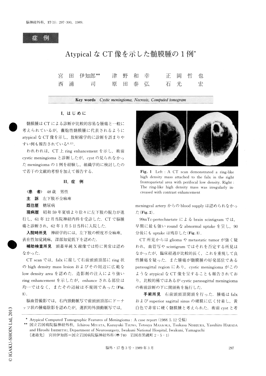
髄膜腫はCTによる診断が比較的容易な腫瘍と一般に考えられているが,嚢胞性髄膜腫に代表されるようにatypicalなCT像を示し,放射線学的に診断を誤まりやすい例も報告されている8,12).
われわれは,CT上ring enhancementを示し,術前cystic meningiomaと診断したが,cystの見られなかったmeningiomaの1例を経験し,組織学的に検討したので若干の文献的考察を加えて報告する.
Diagnosis of intracranial meningiomas with com-puted tomography (CT) is usually easy. However, some authors have reported cases preoperatively misdia-gnosed because of atypical computed tomographic fea-tures. We report a case of non-cystic meningioma pre-senting a ring enhanced mass lesion, with CT scan.
A 48-year-old male was admitted to our hospital on January 5, 1987, because of progressive monoparesis of his left lower limb. This had continued for 11/2 years. Neurological examination revealed monoparesis, hypes-thesia and decreased deep sensation of the left lower limb. X-ray films of the skull appeared normal.
Copyright © 1989, Igaku-Shoin Ltd. All rights reserved.