Japanese
English
- 有料閲覧
PPV購入案内
1,320円 (1,200円+税10%) こちらは、1113ページから1117ページの文献です。購入には医書.jp本会員登録が必要です。
会員登録完了後に改めて上記「購入サイトへ」を選択してください。
または「購入サイトへ」選択後に「購入ログイン」、「新規会員登録」の順に進んでください。
会員登録について詳しくはをご確認ください。
- Abstract 文献概要
- 1ページ目 Look Inside
Ⅰ.はじめに
脊髄血管撮影が脊髄血管腫の診断にきわめて有効な検査法であることはすでに良く知られており,その普及とともに発見率あるいは手術成績が向上してきている.myelogramで蛇行した陰影欠損がみられるときまず考えられるのは血管腫であり,血管撮影によってその性状,大きさ,分布などが確かめられる6,7,10,13).
もしmyelogramでみられた蛇行状の陰影欠損が当を得た血管撮影で造影されないとき,例外的に adhesive arachnoiditis2)あるいはhypertrophic interstitial neuritis of Dejerine-Sottas1)を考慮しなければならないし血管腫自身の血栓形成という報告もある12).
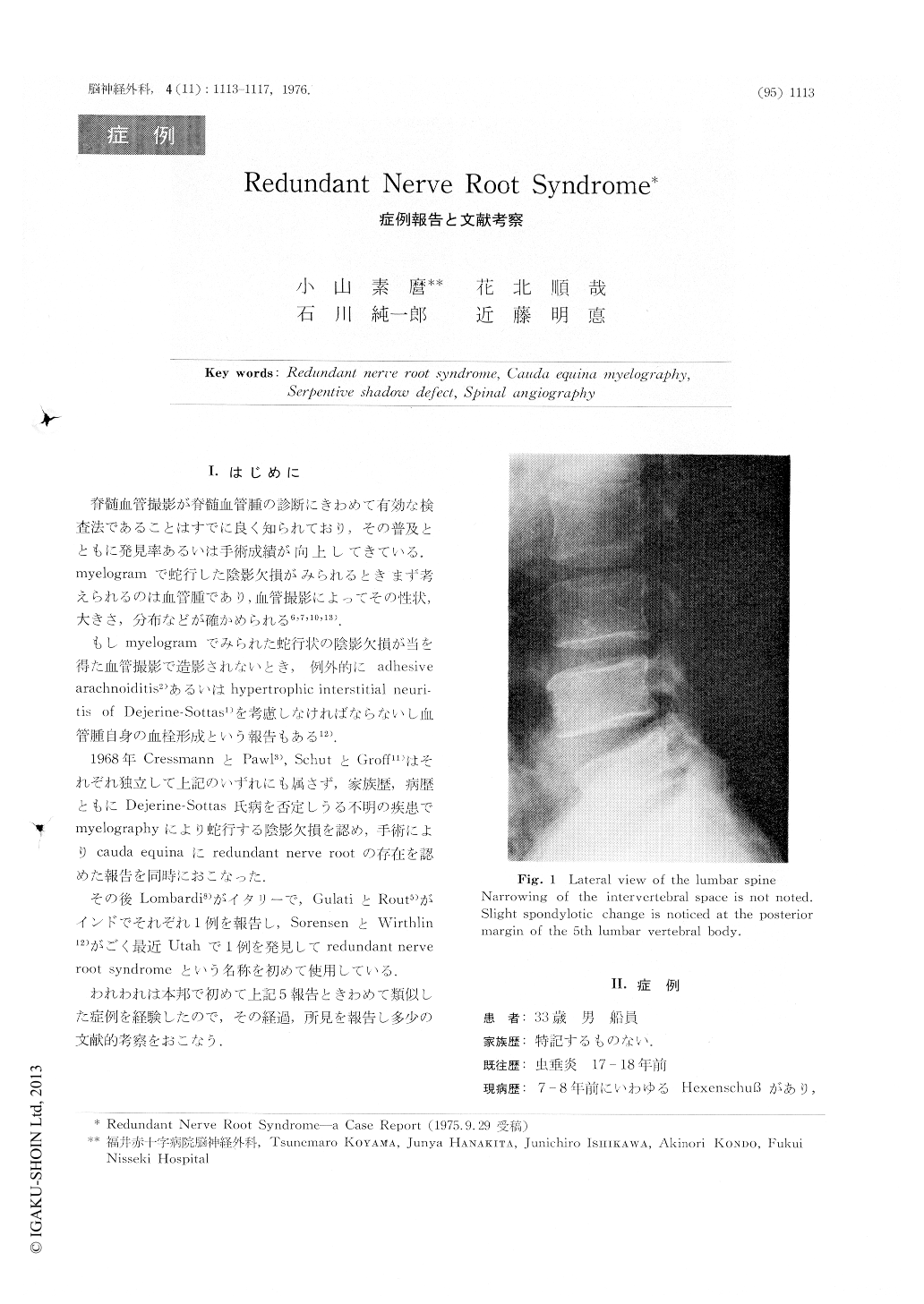
A 33-year-old fisherman was in good health until 1975 when he sustained an injury on his back and experienced an episode of lower back pain radiating to the dorsal aspect of the right leg. About 6 months later, when he was first seen in our clinic his lower back pain was still persisting, and hypesthesia hypoalgesia to pinprick were noted on both L-4, L-5 and S-1 levels. He also complained of impotence and difficulty in urination. A lumbar myelogram with Dimer-X revealed a partial stop of the contrast dye at the level of L3/L4 and also serpentlike shadow defect around the same area.
Copyright © 1976, Igaku-Shoin Ltd. All rights reserved.