Japanese
English
研究
Ependymoblastomaの電子顕微鏡的考察
Electron Microscopic Observations of Ependymoblastoma
平野 朝雄
1
,
松井 孝嘉
2
,
H. M. Zimmerman
3
Asao HIRANO
1
,
Takayoshi MATSUI
2
1Division of Neuropathology, Montefiore Hospital and Medical Center and Professor of Pathology, Albert Einstein College of Medicine
2Division of Neuropathology, Montefiore Hospital and Medical Center and Albert Einstein College of Medicine
3Montefiore Hospital and Medical Center and Professor Emeritus, Albert Einstein College of Medicine
キーワード:
Ependymoblastoma
,
Neural tube
,
Junctional apparatus
,
Rosette
,
Experimental glioma
Keyword:
Ependymoblastoma
,
Neural tube
,
Junctional apparatus
,
Rosette
,
Experimental glioma
pp.467-473
発行日 1975年6月10日
Published Date 1975/6/10
DOI https://doi.org/10.11477/mf.1436200313
- 有料閲覧
PPV購入案内
1,320円 (1,200円+税10%) こちらは、467ページから473ページの文献です。
PPV(ペイ・パー・ビュー)とは、論文(記事)単位で購入・閲覧ができるサービスです。
購入には医書.jp本会員登録が必要です。
会員登録完了後に改めて上記「購入サイトへ」を選択してください。
または「購入サイトへ」選択後に「購入ログイン」、「新規会員登録」の順に進んでください。
会員登録について詳しくはをご確認ください。
購入には医書.jp本会員登録が必要です。
会員登録完了後に改めて上記「購入サイトへ」を選択してください。
または「購入サイトへ」選択後に「購入ログイン」、「新規会員登録」の順に進んでください。
会員登録について詳しくはをご確認ください。
- Abstract 文献概要
- 1ページ目 Look Inside
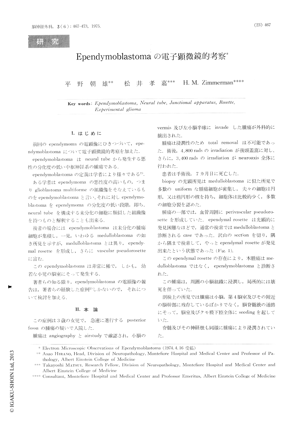
Ⅰ.はじめに
前回のependyniomaの電顕像にひきつづいて,ependymoblastomaについて電子顕微鏡的考察を加えた.
ependymoblastomaはneural tubeから発生する悪性の分化度の低い中枢紳経系の腫瘍である.
The fine structure of an ependymoblastoma was examined and compared to nomal ependyma, ependymoma and to developing neural tube. As in the ependymoma, the tumor cells formed compact masses as well as rosettes and the blood vessel-associated pseudorosettes. In general, the cells appeared primitive but displayed well developed junctional devices in some areas as well as ciliary basal bodies. In many respects, the tumor cells resembled the cells forming the developing neural tube.
Copyright © 1975, Igaku-Shoin Ltd. All rights reserved.