Japanese
English
研究
Ependymaの電子顕微鏡学的考察
Electron Microscopic Observation of Ependyma
平野 朝雄
1
,
松井 孝嘉
2
,
Zimmerman H. M.
3
Asao HIRANO
1
,
Takayoshi MATSUI
2
1Division of Neuropathology, Montefiore Hospital and Medical Center and Professor of Pathology, Albert Einstein College of Medicine
2Division of Neuropathology, Montefiore Hospital and Medical Center and Albert Einstein College of Medicine
3Consultant, Montefiore Hospital and Medical Center and Professor Emeritus, Albert Einstein College of Medicine
キーワード:
Ependyma
,
Cilia
,
Microvilli
,
Basement membrane
,
Filament
Keyword:
Ependyma
,
Cilia
,
Microvilli
,
Basement membrane
,
Filament
pp.237-244
発行日 1975年3月10日
Published Date 1975/3/10
DOI https://doi.org/10.11477/mf.1436200280
- 有料閲覧
PPV購入案内
1,320円 (1,200円+税10%) こちらは、237ページから244ページの文献です。
PPV(ペイ・パー・ビュー)とは、論文(記事)単位で購入・閲覧ができるサービスです。
購入には医書.jp本会員登録が必要です。
会員登録完了後に改めて上記「購入サイトへ」を選択してください。
または「購入サイトへ」選択後に「購入ログイン」、「新規会員登録」の順に進んでください。
会員登録について詳しくはをご確認ください。
購入には医書.jp本会員登録が必要です。
会員登録完了後に改めて上記「購入サイトへ」を選択してください。
または「購入サイトへ」選択後に「購入ログイン」、「新規会員登録」の順に進んでください。
会員登録について詳しくはをご確認ください。
- Abstract 文献概要
- 1ページ目 Look Inside
Ⅰ.緒論
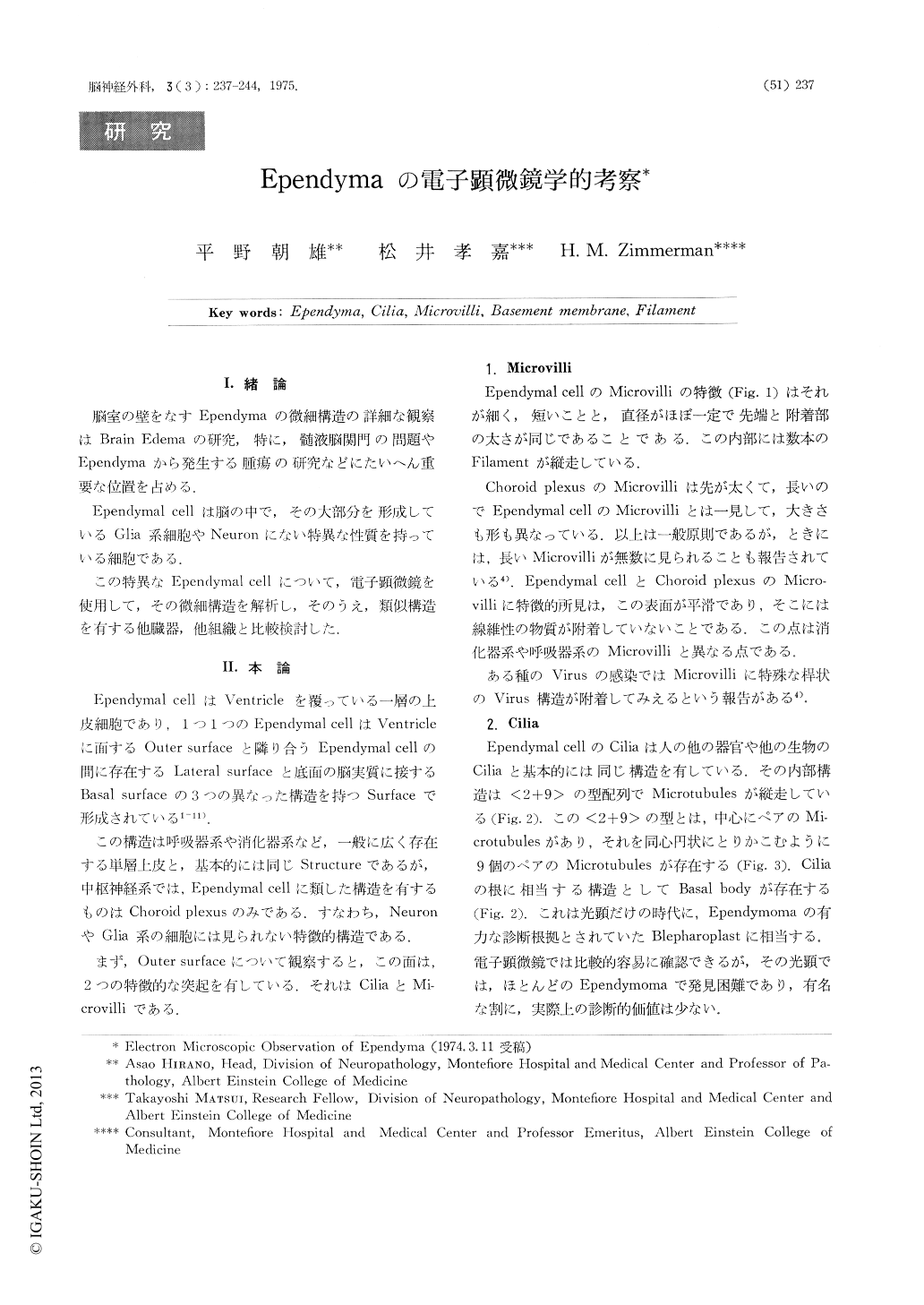
脳室の壁をなすEpendymaの微細構造の詳細な観察はBrain Edemaの研究,特に,髄液脳関門の問題やEpendymaから発生する腫瘍の研究などにたいへん重要な位置を占める.
Ependymal cellは脳の中で,その大部分を形成しているGlia系細胞やNeuronにない特異な性質を持っている細胞である.
The fine structure of the normal ependymal cell has been described. The ependymal cells form a closely knit single layer lining the ventricles. They are bordered on one side by the ventricular lumen and by the neuropil on the basal surface.
The luminal surface is characterized by microvilfi and cilia. The former differ from those seen in the respiratory tract or intestine in that they have a smooth surface and are devoid of a coating material. They differ from the microvilli of the choroid plexus by being straight and relatively stubby.
Copyright © 1975, Igaku-Shoin Ltd. All rights reserved.