Japanese
English
症例
線維化や血管腫像を主体とした小脳腫瘍—小脳腫瘍の終末的病像
Cerebellar Tumors Manifesting Fibrosis and A Hemangiomatous Pattern
武田 文和
1
,
田村 勝
1
,
八木 久男
1
,
深町 彰
1
Fumikazu TAKEDA
1
,
Masaru TAMURA
1
,
Hisao YAGI
1
,
Akira FUKAMACHI
1
1群馬大学脳神経外科
1Department of Neurosurgery, School of Medicine, Gunma University
キーワード:
Cerebellar tumor
,
Hemorrhage
,
Necrosis
,
Fibrosis
,
Sclerosing hemangioma
Keyword:
Cerebellar tumor
,
Hemorrhage
,
Necrosis
,
Fibrosis
,
Sclerosing hemangioma
pp.559-564
発行日 1974年8月10日
Published Date 1974/8/10
DOI https://doi.org/10.11477/mf.1436200215
- 有料閲覧
PPV購入案内
1,320円 (1,200円+税10%) こちらは、559ページから564ページの文献です。
PPV(ペイ・パー・ビュー)とは、論文(記事)単位で購入・閲覧ができるサービスです。
購入には医書.jp本会員登録が必要です。
会員登録完了後に改めて上記「購入サイトへ」を選択してください。
または「購入サイトへ」選択後に「購入ログイン」、「新規会員登録」の順に進んでください。
会員登録について詳しくはをご確認ください。
購入には医書.jp本会員登録が必要です。
会員登録完了後に改めて上記「購入サイトへ」を選択してください。
または「購入サイトへ」選択後に「購入ログイン」、「新規会員登録」の順に進んでください。
会員登録について詳しくはをご確認ください。
- Abstract 文献概要
- 1ページ目 Look Inside
Ⅰ.はじめに
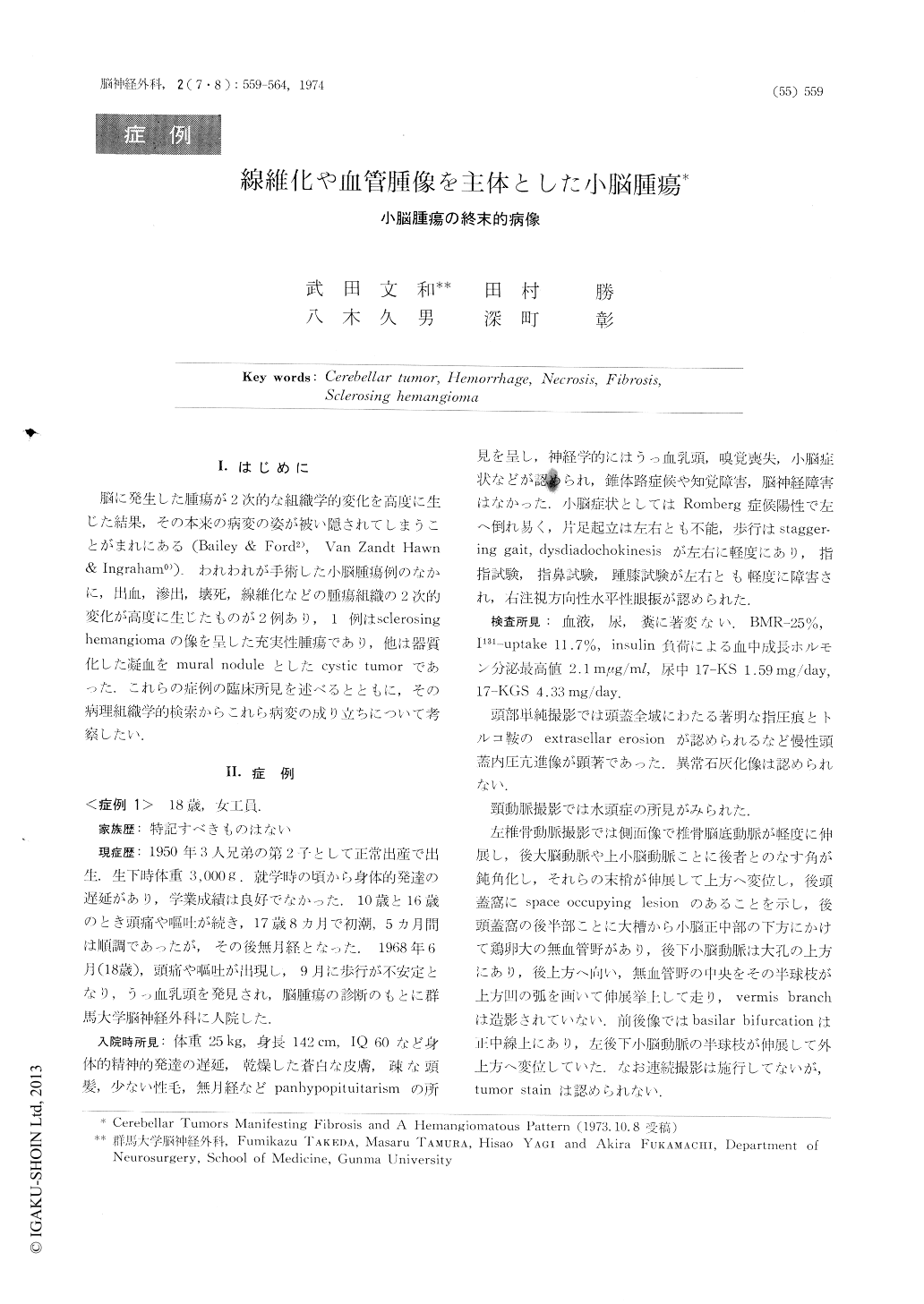
脳に発生した腫瘍が2次的な組織学的変化を高度に生じた結果,その本来の病変の姿が被い隠されてしまうことがまれにある(Bailey & Ford2),Van Zandt Hawn & Ingraham0)).われわれが手術した小脳腫瘍例のなかに,出血,滲出,壊死,線維化などの腫瘍組織の2次的変化が高度に生じたものが2例あり,1例はsclerosing hemangiomaの像を呈した充実性腫瘍であり,他は器質化した凝血をmural noduleとしたcystic tumorであった.これらの症例の臨床所見を述べるとともに,その病理組織学的検索からこれら病変の成り立ちについて考察したい.
Two cases with cerebellar tumor were reported in which secondary histological sequences of the tumor tissues were so marked as to mark completely the essential histological features of the lesions. Histological examination suggested that those two tumors might represent the outcome of progressive tissue changes occurring in cere bellar hemangioma, fibroma, astrocytoms or hemangioblastoma.
Case 1. The patient was an 18 yearold female.
Copyright © 1974, Igaku-Shoin Ltd. All rights reserved.