Japanese
English
- 有料閲覧
PPV購入案内
1,320円 (1,200円+税10%) こちらは、127ページから133ページの文献です。購入には医書.jp本会員登録が必要です。
会員登録完了後に改めて上記「購入サイトへ」を選択してください。
または「購入サイトへ」選択後に「購入ログイン」、「新規会員登録」の順に進んでください。
会員登録について詳しくはをご確認ください。
- Abstract 文献概要
- 1ページ目 Look Inside
- 参考文献 Reference
Ⅰ.はじめに
胸部大動脈解離の代表的な症状が胸背部痛であることはよく知られており,その場合の診断は容易であるが,10~55%の症例では胸背部痛がないとされ,その場合の診断は容易ではない4).また,胸部大動脈解離では脳,脊髄,末梢神経由来などの神経症状が6~42%に出現するとされているが,そのうち意識障害で発症した場合は病歴が聴取できず,診断が困難である4,11,14).
われわれはshock状態により意識障害を生じたもののMRIにて脳に異常を認めず,診断が困難であった胸部大動脈解離の1例を経験した.今後の診療の一助となり得ると考え,本症例を報告する.
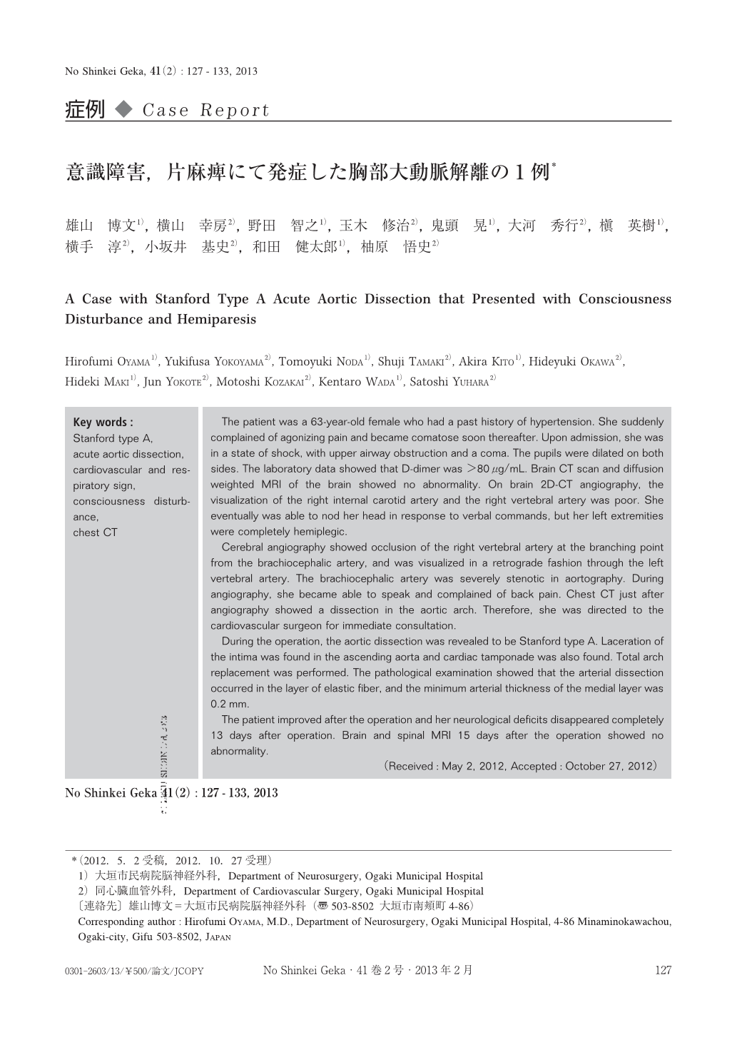
The patient was a 63-year-old female who had a past history of hypertension. She suddenly complained of agonizing pain and became comatose soon thereafter. Upon admission, she was in a state of shock, with upper airway obstruction and a coma. The pupils were dilated on both sides. The laboratory data showed that D-dimer was >80μg/mL. Brain CT scan and diffusion weighted MRI of the brain showed no abnormality. On brain 2D-CT angiography, the visualization of the right internal carotid artery and the right vertebral artery was poor. She eventually was able to nod her head in response to verbal commands, but her left extremities were completely hemiplegic.
Cerebral angiography showed occlusion of the right vertebral artery at the branching point from the brachiocephalic artery, and was visualized in a retrograde fashion through the left vertebral artery. The brachiocephalic artery was severely stenotic in aortography. During angiography, she became able to speak and complained of back pain. Chest CT just after angiography showed a dissection in the aortic arch. Therefore, she was directed to the cardiovascular surgeon for immediate consultation.
During the operation, the aortic dissection was revealed to be Stanford type A. Laceration of the intima was found in the ascending aorta and cardiac tamponade was also found. Total arch replacement was performed. The pathological examination showed that the arterial dissection occurred in the layer of elastic fiber, and the minimum arterial thickness of the medial layer was 0.2mm.
The patient improved after the operation and her neurological deficits disappeared completely 13 days after operation. Brain and spinal MRI 15 days after the operation showed no abnormality.
Copyright © 2013, Igaku-Shoin Ltd. All rights reserved.