Japanese
English
- 有料閲覧
- Abstract 文献概要
- 1ページ目 Look Inside
- 参考文献 Reference
Ⅰ.はじめに
中枢神経原発悪性リンパ腫(primary central nervous system lymphoma:PCNSL)の原発性脳腫瘍に占める割合は約3%と比較的稀であるが,近年増加傾向にある12).高齢者に好発することや臓器移植後やacquired immunodeficiency syndrome(AIDS)のような免疫不全状態での発生が知られており,発生率増加の一因と考えられている.
治療に関しては,high-dose methotrexate(HD-MTX)とそれに続く全脳照射(whole brain radiotherapy:WBRT)の併用療法の有効性が報告され,生存期間中央値は30~40カ月と延長し,長期生存する症例も多数認められている16).PCNSLは臨床的に症状が出現してからの進行が速いことが特徴で,早期の診断と治療開始がさらなる生存期間の延長には必要である.PCNSLに特徴的な症状・神経所見はなく,magnetic resonance imaging(MRI)による画像診断が有用なことが多い1,3,11).しかし,非典型的な形態学的画像所見を呈するPCNSLも多数存在し4),診断が遅れて剖検ではじめてPCNSLと診断されることもしばしばある4).
2-[18F]-fluoro-2-deoxy-D-glucose(FDG)を用いた陽電子断層撮影(positron emission tomography:PET)は,腫瘍の悪性度をブドウ糖代謝の面から評価することのできる分子イメージングである.一般的に腫瘍細胞においては,悪性度の高いものほど細胞密度が高く,またグルコース輸送体膜蛋白(glucose transporter:GLUT)が過剰発現し,細胞内のhexokinaze活性も亢進しており腫瘍へのFDG集積は増加する.PCNSLにおけるFDG-PETの役割は確立されたものではないが,近年の臨床研究においてその有用性が報告されており6,10,15,17),われわれもPCNSLが疑われる症例には全例にFDG-PET検査を行ってきた.その中で,典型的画像所見を呈する症例ではFDG-PETは画像診断として非常に有用であり6,15),鑑別診断や病理組織学的診断が行えない症例への応用も期待される一方で,非典型的画像所見を呈する症例ではその有用性に限界があることがわかってきた7).全国的なPET装置の普及に伴いPCNSLの診断にデリバリーによるFDGを用いたPET検査を行う機会が増加することが予想される.今回,われわれが経験した全症例をまとめることで,PCNSLの診断におけるFDG-PETの有用性と問題点について文献的考察を加えて報告する.
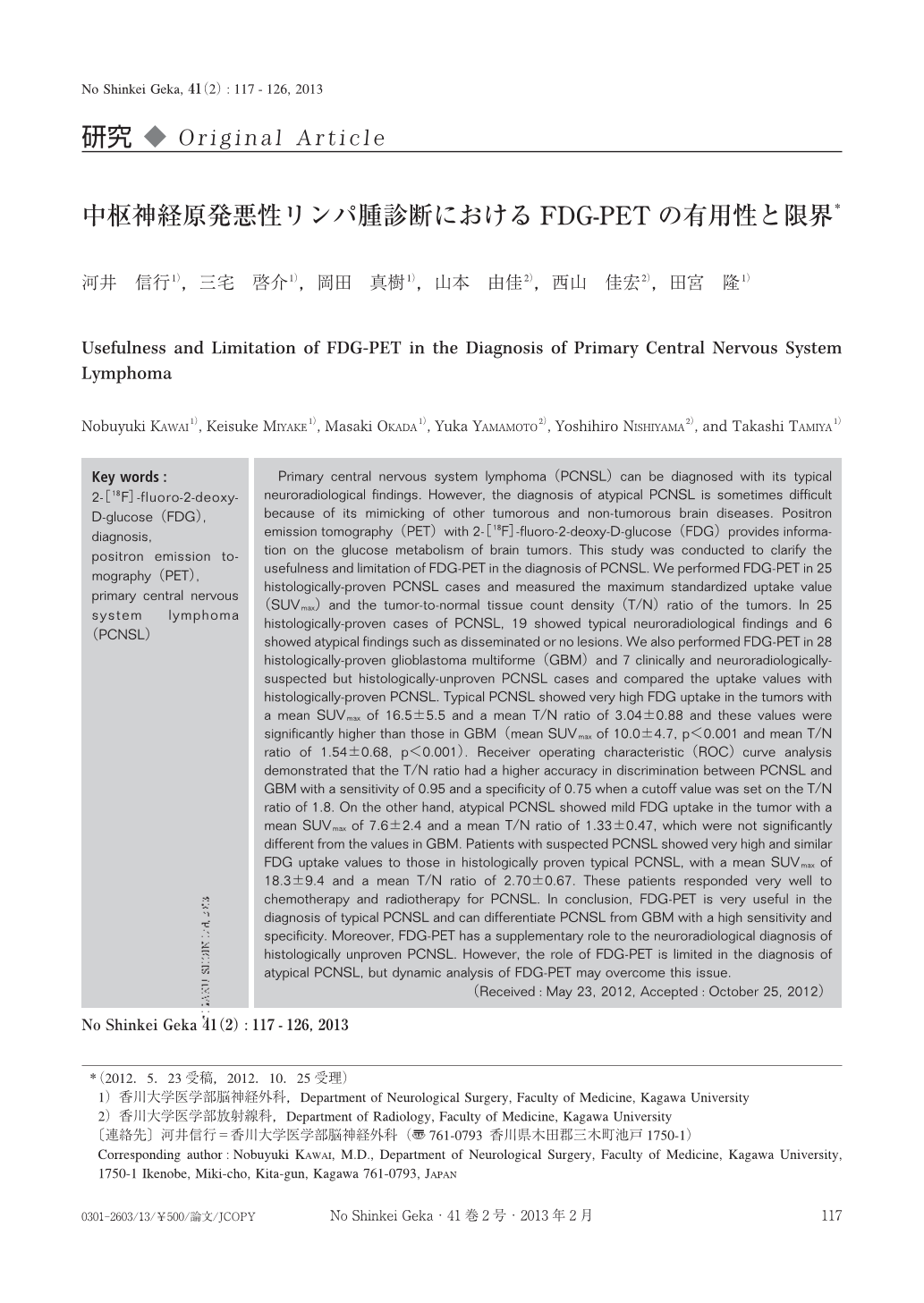
Primary central nervous system lymphoma(PCNSL)can be diagnosed with its typical neuroradiological findings. However, the diagnosis of atypical PCNSL is sometimes difficult because of its mimicking of other tumorous and non-tumorous brain diseases. Positron emission tomography(PET)with 2-[18F]-fluoro-2-deoxy-D-glucose(FDG)provides information on the glucose metabolism of brain tumors. This study was conducted to clarify the usefulness and limitation of FDG-PET in the diagnosis of PCNSL. We performed FDG-PET in 25 histologically-proven PCNSL cases and measured the maximum standardized uptake value(SUVmax)and the tumor-to-normal tissue count density(T/N)ratio of the tumors. In 25 histologically-proven cases of PCNSL, 19 showed typical neuroradiological findings and 6 showed atypical findings such as disseminated or no lesions. We also performed FDG-PET in 28 histologically-proven glioblastoma multiforme(GBM)and 7 clinically and neuroradiologically-suspected but histologically-unproven PCNSL cases and compared the uptake values with histologically-proven PCNSL. Typical PCNSL showed very high FDG uptake in the tumors with a mean SUVmax of 16.5±5.5 and a mean T/N ratio of 3.04±0.88 and these values were significantly higher than those in GBM(mean SUVmax of 10.0±4.7, p<0.001 and mean T/N ratio of 1.54±0.68, p<0.001). Receiver operating characteristic(ROC)curve analysis demonstrated that the T/N ratio had a higher accuracy in discrimination between PCNSL and GBM with a sensitivity of 0.95 and a specificity of 0.75 when a cutoff value was set on the T/N ratio of 1.8. On the other hand, atypical PCNSL showed mild FDG uptake in the tumor with a mean SUVmax of 7.6±2.4 and a mean T/N ratio of 1.33±0.47, which were not significantly different from the values in GBM. Patients with suspected PCNSL showed very high and similar FDG uptake values to those in histologically proven typical PCNSL, with a mean SUVmax of 18.3±9.4 and a mean T/N ratio of 2.70±0.67. These patients responded very well to chemotherapy and radiotherapy for PCNSL. In conclusion, FDG-PET is very useful in the diagnosis of typical PCNSL and can differentiate PCNSL from GBM with a high sensitivity and specificity. Moreover, FDG-PET has a supplementary role to the neuroradiological diagnosis of histologically unproven PCNSL. However, the role of FDG-PET is limited in the diagnosis of atypical PCNSL, but dynamic analysis of FDG-PET may overcome this issue.

Copyright © 2013, Igaku-Shoin Ltd. All rights reserved.


