Japanese
English
- 有料閲覧
PPV購入案内
1,320円 (1,200円+税10%) こちらは、991ページから1000ページの文献です。購入には医書.jp本会員登録が必要です。
会員登録完了後に改めて上記「購入サイトへ」を選択してください。
または「購入サイトへ」選択後に「購入ログイン」、「新規会員登録」の順に進んでください。
会員登録について詳しくはをご確認ください。
- Abstract 文献概要
- 1ページ目 Look Inside
- 参考文献 Reference
Ⅰ.はじめに
脳梗塞の2~3割は,頸動脈病変が原因と考えられている.しかし,その原因である血管壁のプラーク性状についての評価は十分になされていない.血管壁はミリ単位という微細な領域であることやプラーク内組織の多様性(新生血管,壁内出血,脂質成分,石灰化,線維化)などから,今まで信頼性のある臨床的評価を下せる検査法がなかったことに原因がある.今後,臨床的にはプラークの評価が可能な検査法が必須で,そのような検査法を通じてプラーク病変の進展機序や病態が解明され,脳虚血発作の予防や手術リスク軽減に寄与していくものと思われる1,7).
今回は,超音波検査のmodalityの1つであるharmonic imaging法(以後HI法)の下で超音波造影剤を使用し,病理学的に観察されるプラーク内新生血管(以後NV)による血液灌流について検討し,さらには3次元的real timeプラーク内血液灌流所見も提示することで,若干の文献考察を加え報告する.
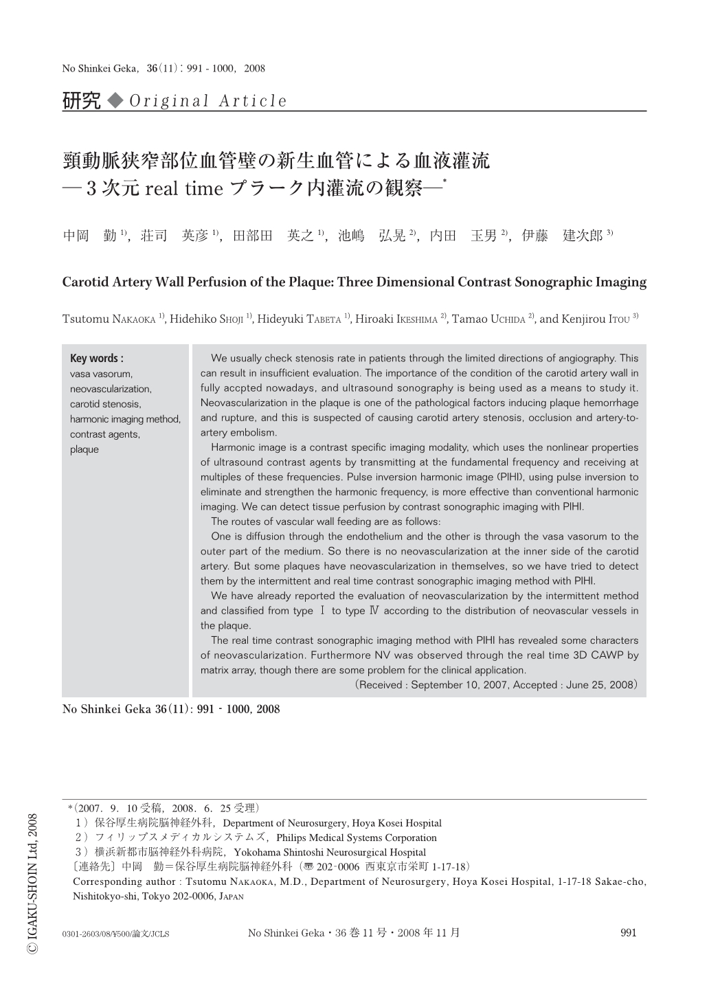
We usually check stenosis rate in patients through the limited directions of angiography. This can result in insufficient evaluation. The importance of the condition of the carotid artery wall in fully accpted nowadays, and ultrasound sonography is being used as a means to study it. Neovascularization in the plaque is one of the pathological factors inducing plaque hemorrhage and rupture, and this is suspected of causing carotid artery stenosis, occlusion and artery-to-artery embolism.
Harmonic image is a contrast specific imaging modality, which uses the nonlinear properties of ultrasound contrast agents by transmitting at the fundamental frequency and receiving at multiples of these frequencies. Pulse inversion harmonic image (PIHI), using pulse inversion to eliminate and strengthen the harmonic frequency, is more effective than conventional harmonic imaging. We can detect tissue perfusion by contrast sonographic imaging with PIHI.
The routes of vascular wall feeding are as follows:
One is diffusion through the endothelium and the other is through the vasa vasorum to the outer part of the medium. So there is no neovascularization at the inner side of the carotid artery. But some plaques have neovascularization in themselves, so we have tried to detect them by the intermittent and real time contrast sonographic imaging method with PIHI.
We have already reported the evaluation of neovascularization by the intermittent method and classified from type Ⅰ to type Ⅳ according to the distribution of neovascular vessels in the plaque.
The real time contrast sonographic imaging method with PIHI has revealed some characters of neovascularization. Furthermore NV was observed through the real time 3D CAWP by matrix array, though there are some problem for the clinical application.
Copyright © 2008, Igaku-Shoin Ltd. All rights reserved.