Japanese
English
- 有料閲覧
PPV購入案内
1,320円 (1,200円+税10%) こちらは、605ページから610ページの文献です。購入には医書.jp本会員登録が必要です。
会員登録完了後に改めて上記「購入サイトへ」を選択してください。
または「購入サイトへ」選択後に「購入ログイン」、「新規会員登録」の順に進んでください。
会員登録について詳しくはをご確認ください。
- Abstract 文献概要
- 1ページ目 Look Inside
- 参考文献 Reference
Ⅰ.は じ め に
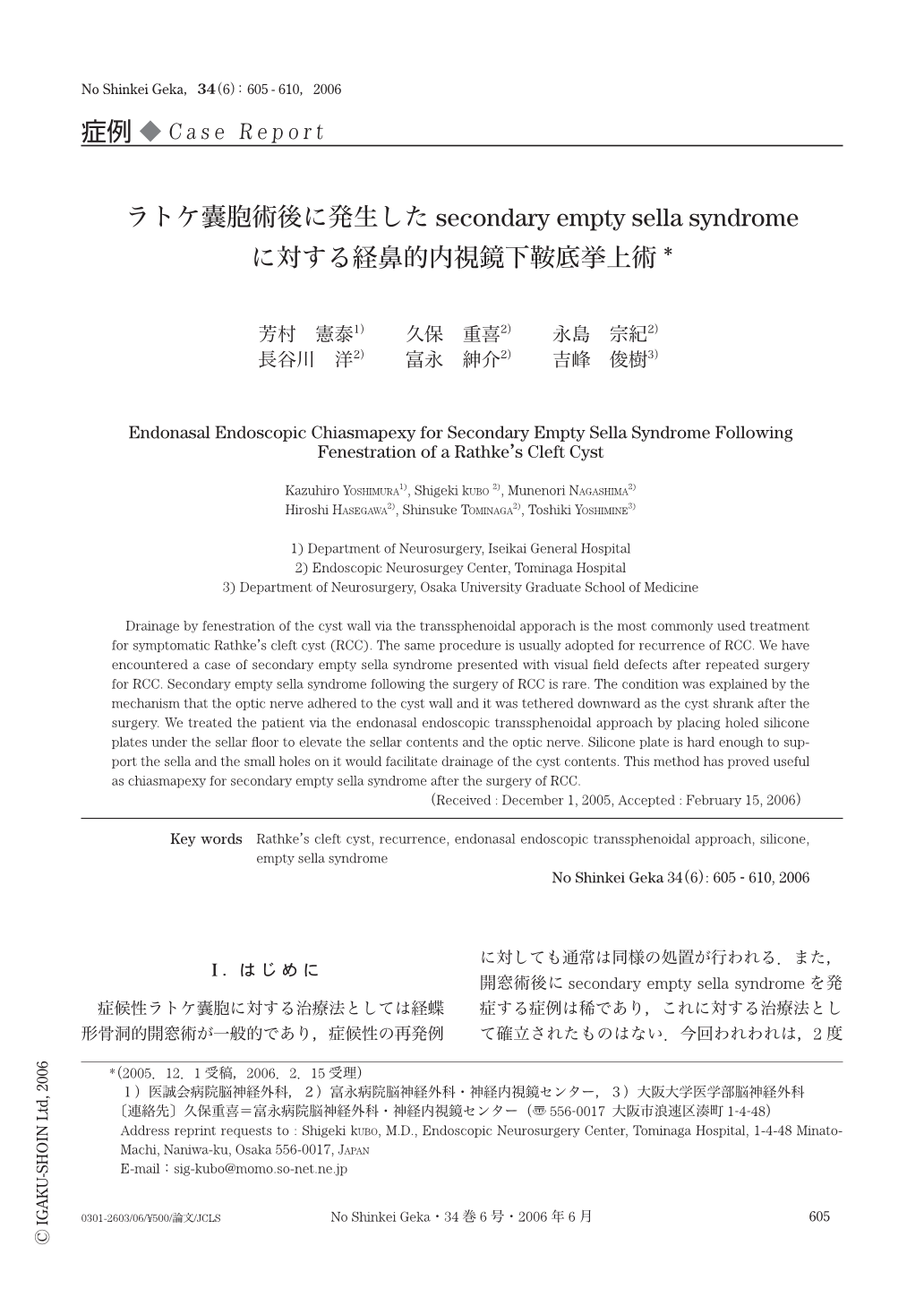
症候性ラトケ嚢胞に対する治療法としては経蝶形骨洞的開窓術が一般的であり,症候性の再発例に対しても通常は同様の処置が行われる.また,開窓術後にsecondary empty sella syndromeを発症する症例は稀であり,これに対する治療法として確立されたものはない.今回われわれは,2度の開窓術後に視野障害で発症したsecondary empty sella syndromeの症例を経験した.Secondary empty sella syndromeの発症機序に対して考察を加え,治療法に関してはわれわれの工夫を報告する.
Drainage by fenestration of the cyst wall via the transsphenoidal apporach is the most commonly used treatment for symptomatic Rathke's cleft cyst (RCC). The same procedure is usually adopted for recurrence of RCC. We have encountered a case of secondary empty sella syndrome presented with visual field defects after repeated surgery for RCC. Secondary empty sella syndrome following the surgery of RCC is rare. The condition was explained by the mechanism that the optic nerve adhered to the cyst wall and it was tethered downward as the cyst shrank after the surgery. We treated the patient via the endonasal endoscopic transsphenoidal approach by placing holed silicone plates under the sellar floor to elevate the sellar contents and the optic nerve. Silicone plate is hard enough to support the sella and the small holes on it would facilitate drainage of the cyst contents. This method has proved useful as chiasmapexy for secondary empty sella syndrome after the surgery of RCC.
Copyright © 2006, Igaku-Shoin Ltd. All rights reserved.