Japanese
English
- 有料閲覧
PPV購入案内
1,320円 (1,200円+税10%) こちらは、417ページから420ページの文献です。購入には医書.jp本会員登録が必要です。
会員登録完了後に改めて上記「購入サイトへ」を選択してください。
または「購入サイトへ」選択後に「購入ログイン」、「新規会員登録」の順に進んでください。
会員登録について詳しくはをご確認ください。
- Abstract 文献概要
- 1ページ目 Look Inside
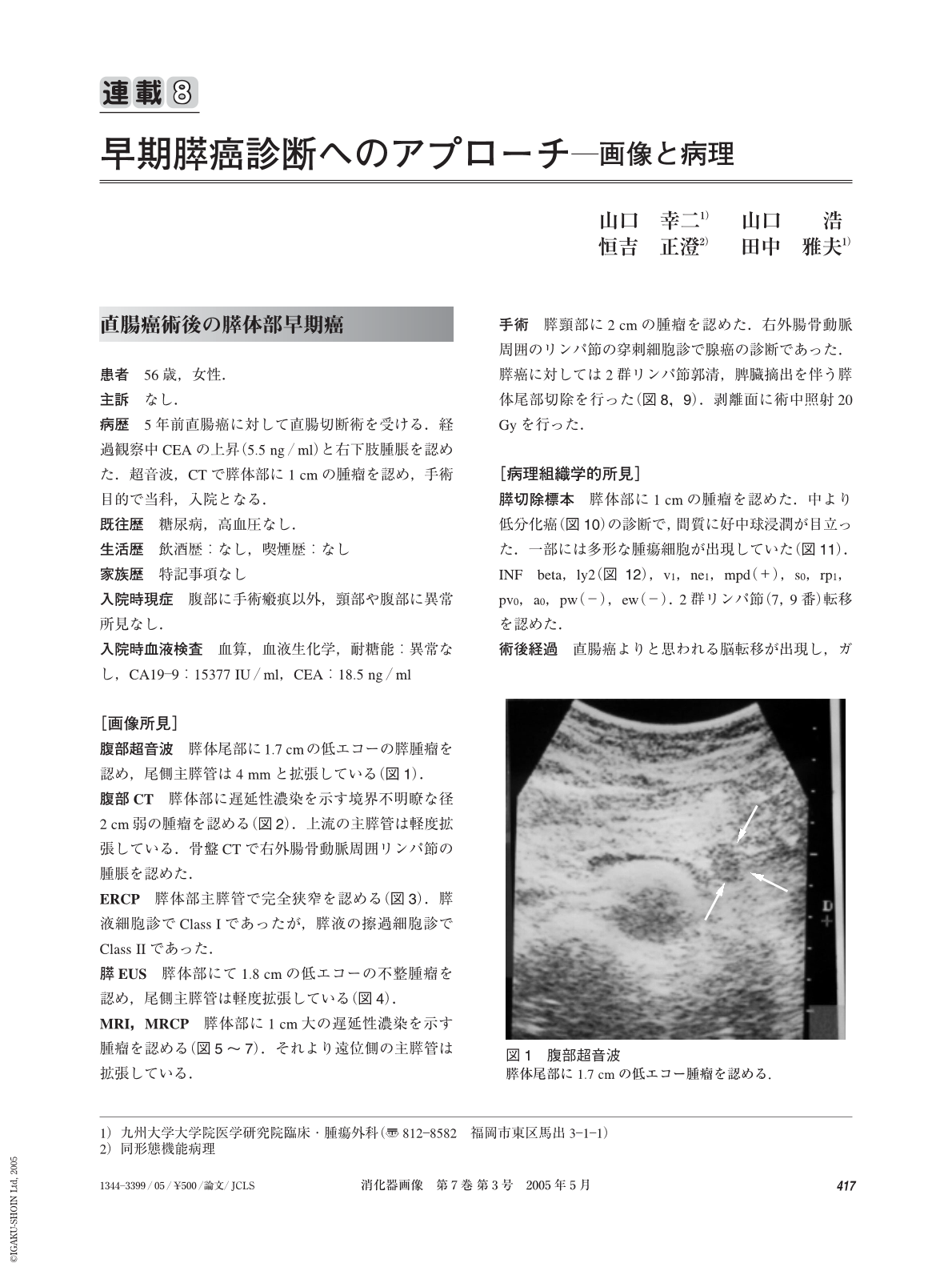
患者 56歳,女性.
主訴 なし.
病歴 5年前直腸癌に対して直腸切断術を受ける.経過観察中CEAの上昇(5.5 ng/ml)と右下肢腫脹を認めた.超音波,CTで膵体部に1 cmの腫瘤を認め,手術目的で当科,入院となる.
既往歴 糖尿病,高血圧なし.
A 56―year-old woman underwent Miles’ operation for rectal carcinoma five years ago. She noticed swelling of right lower extremity and serum CEA level was elevated. Ultrasonography showed a pancreatic body mass, measuring 1 cm, and she came to us for surgical resection. Serum level of CA19―9 was elevated to 15,377 IU/ml and CEA 18.5 ng/ml. Computed tomography and magnetic resonance imaging also showed a pancreatic body mass with delayed enhancement and dilatation of the upper stream pancreatic ducts. Lymph node swelling along the external iliac artery was evident. Endoscopic retrograde pancreatography showed a complete obstruction of the main pancreatic duct. Distal pancreatectomy and splenectomy was done with D2 lymph node dissection. Intraoperative radiation therapy(20 Gy)was also done. Biopsy of lymph node around the right external iliac artery showed metastasis by cytology. Histological examination showed a moderately to poorly differentiated adenocarcinoma, measuring 1.5 cm. Metastasis was not evident in level 2 lymph nodes. After operation, brain metastasis from rectal cancer developed and died three months after operation.
(Shokakigazo 2005 ; 7 : 417―420)
Copyright © 2005, Igaku-Shoin Ltd. All rights reserved.