Japanese
English
- 有料閲覧
PPV購入案内
1,320円 (1,200円+税10%) こちらは、467ページから474ページの文献です。購入には医書.jp本会員登録が必要です。
会員登録完了後に改めて上記「購入サイトへ」を選択してください。
または「購入サイトへ」選択後に「購入ログイン」、「新規会員登録」の順に進んでください。
会員登録について詳しくはをご確認ください。
- Abstract 文献概要
- 1ページ目 Look Inside
- 参考文献 Reference
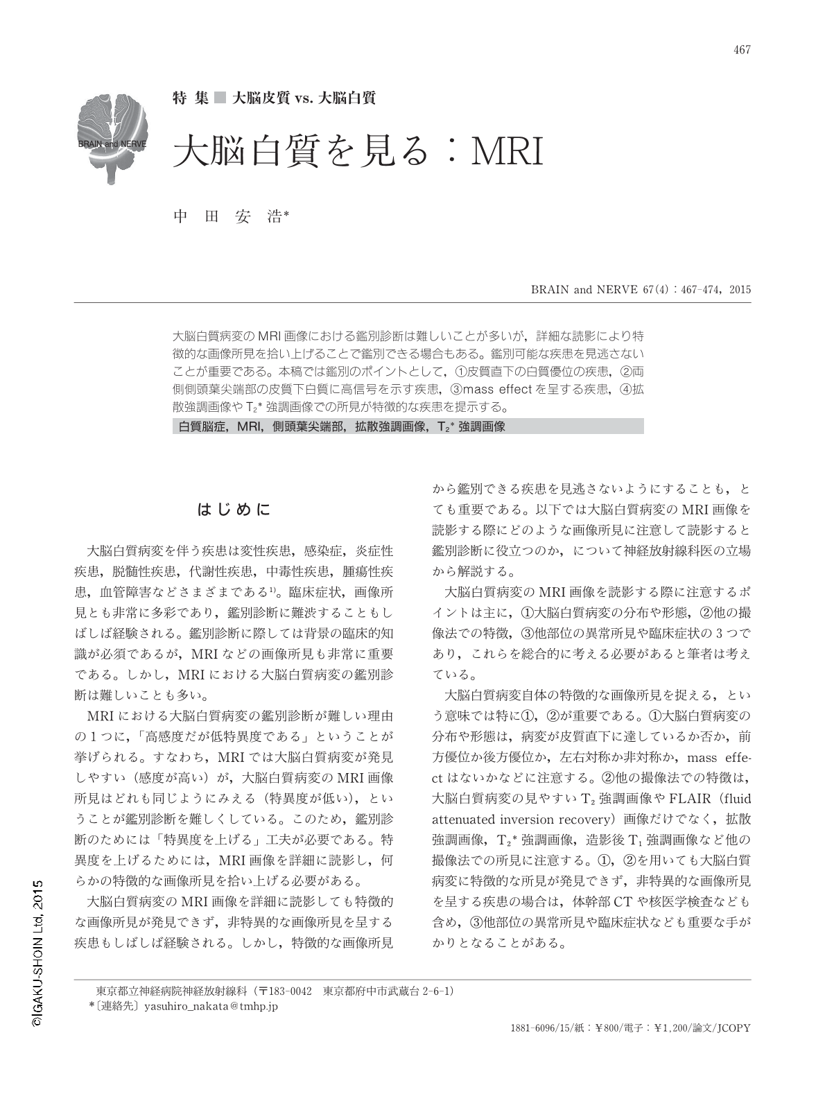
大脳白質病変のMRI画像における鑑別診断は難しいことが多いが,詳細な読影により特徴的な画像所見を拾い上げることで鑑別できる場合もある。鑑別可能な疾患を見逃さないことが重要である。本稿では鑑別のポイントとして,①皮質直下の白質優位の疾患,②両側側頭葉尖端部の皮質下白質に高信号を示す疾患,③mass effectを呈する疾患,④拡散強調画像やT2*強調画像での所見が特徴的な疾患を提示する。
Abstract
It is often difficult to make a differential diagnosis of cerebral white-matter lesions on magnetic resonance images (MRI), because imaging findings are non-specific. However, it is possible to make a correct diagnosis of some kinds of cerebral white-matter lesions upon a detailed analysis of MRI. In analyzing MRI of cerebral white matter lesions, the localization and shape of white-matter lesions are important factors to make a differential diagnosis. Other images such as diffusion-weighted or T2-star weighted images are sometimes also useful for making such a diagnosis. In this manuscript, I describe how to read MRI of cerebral white-matter lesions, and present some educational cases.
Copyright © 2015, Igaku-Shoin Ltd. All rights reserved.