Japanese
English
- 有料閲覧
PPV購入案内
1,320円 (1,200円+税10%) こちらは、249ページから260ページの文献です。購入には医書.jp本会員登録が必要です。
会員登録完了後に改めて上記「購入サイトへ」を選択してください。
または「購入サイトへ」選択後に「購入ログイン」、「新規会員登録」の順に進んでください。
会員登録について詳しくはをご確認ください。
- Abstract 文献概要
- 1ページ目 Look Inside
- 参考文献 Reference
- サイト内被引用 Cited by
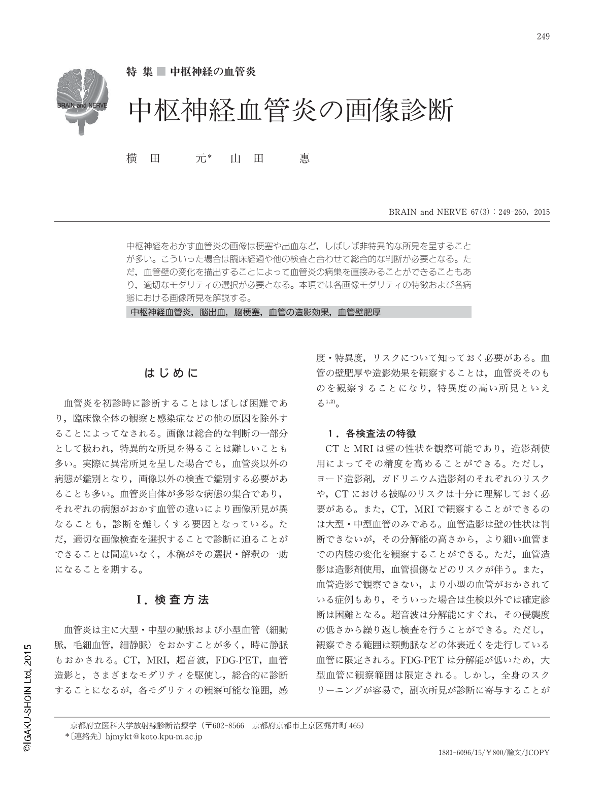
中枢神経をおかす血管炎の画像は梗塞や出血など,しばしば非特異的な所見を呈することが多い。こういった場合は臨床経過や他の検査と合わせて総合的な判断が必要となる。ただ,血管壁の変化を描出することによって血管炎の病巣を直接みることができることもあり,適切なモダリティの選択が必要となる。本項では各画像モダリティの特徴および各病態における画像所見を解説する。
Abstract
Vasculitis involving the central nervous system presents with infarction and hemorrhage, which are often nonspecific findings. Laboratory examinations are essential for diagnosis of vasculitis in addition to comprehensive and systematic review of the clinical course. Although most findings tend to be nonspecific, enhancement and thickening of the vascular wall indicate vasculitis. Visualization of the vascular wall requires selection of the appropriate imaging modality and mode of image acquisition. Contrast-enhanced CT, MRI, and FDG-PET are useful for visualizing large vessel vasculitis, while CT, MRI, and angiography are effective for medium vessel vasculitis. The use of ultrasound is limited to evaluating vessels on the body surface. Although relatively thick vessels can be demonstrated by angiography, complete survey of small vessels is difficult. Here, we summarize the characteristics of each imaging modality and imaging findings of typical vasculitides—Takayasu arteritis, giant cell arteritis, ANCA-associated vasculitis, Behçet's disease, primary angiitis of the CNS, and vasculitis associated with systemic disease. Differential diagnoses are also shown, including infective endocarditis, tuberculous meningitis, Ehlers-Danlos syndrome, and reversible cerebral vasoconstriction syndrome.
Copyright © 2015, Igaku-Shoin Ltd. All rights reserved.