Japanese
English
- 有料閲覧
PPV購入案内
1,320円 (1,200円+税10%) こちらは、547ページから553ページの文献です。購入には医書.jp本会員登録が必要です。
会員登録完了後に改めて上記「購入サイトへ」を選択してください。
または「購入サイトへ」選択後に「購入ログイン」、「新規会員登録」の順に進んでください。
会員登録について詳しくはをご確認ください。
- Abstract 文献概要
- 1ページ目 Look Inside
- 参考文献 Reference
はじめに
現在,脳虚血患者に対する脳血流の評価は,SPECTや脳血管撮影などの方法で行われている。しかしこれらの従来の検査方法は,解決すべきさまざまな問題点がある。すなわち,①検査時間が長い,②検査中に患者の拘束度が高い,③リアルタイムに測定できない,④患者を病室から検査室まで移動させなければならず重症患者では検査し難い,⑤緊急時に即座に対応できない,⑥設備が大掛かりで高額である,といった点が考えられる。
近年臨床的に使用されるようになった光トポグラフィーは,その特徴として,①簡便であること,②被験者に対して無侵襲,非拘束的であること,③リアルタイムにかつ持続的に測定可能であること,④可搬性でありベッドサイドでも測定可能であること,⑤速やかに検査が可能であること,などが挙げられる1,2)。
われわれは,光トポグラフィーのこれらの特徴を利用して,脳虚血患者の脳血流状態をリアルタイムに,場所を選ばず,無侵襲で測定解析することができれば従来の検査方法の欠点を補い,脳虚血の診断や治療のうえで迅速かつ的確な対応ができると考え,光トポグラフィーによる脳虚血の評価方法を開発,検討した。
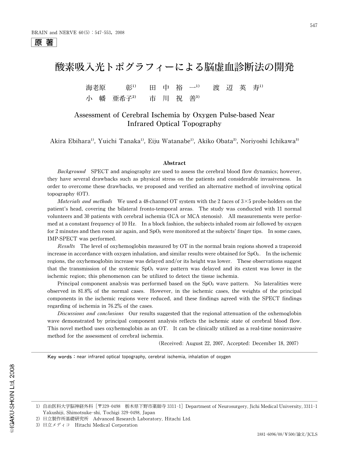
Abstract
Background SPECT and angiography are used to assess the cerebral blood flow dynamics; however, they have several drawbacks such as physical stress on the patients and considerable invasiveness. In order to overcome these drawbacks, we proposed and verified an alternative method of involving optical topography (OT).
Materials and methods We used a 48-channel OT system with the 2 faces of 3×5 probe-holders on the patient's head, covering the bilateral fronto-temporal areas. The study was conducted with 11 normal volunteers and 30 patients with cerebral ischemia (ICA or MCA stenosis). All measurements were performed at a constant frequency of 10 Hz. In a block fashion, the subjects inhaled room air followed by oxygen for 2 minutes and then room air again, and SpO2 were monitored at the subjects' finger tips. In some cases, IMP-SPECT was performed.
Results The level of oxyhemoglobin measured by OT in the normal brain regions showed a trapezoid increase in accordance with oxygen inhalation, and similar results were obtained for SpO2. In the ischemic regions, the oxyhemoglobin increase was delayed and/or its height was lower. These observations suggest that the transmission of the systemic SpO2 wave pattern was delayed and its extent was lower in the ischemic region; this phenomenon can be utilized to detect the tissue ischemia.
Principal component analysis was performed based on the SpO2 wave pattern. No lateralities were observed in 81.8% of the normal cases. However, in the ischemic cases, the weights of the principal components in the ischemic regions were reduced, and these findings agreed with the SPECT findings regarding of ischemia in 76.2% of the cases.
Discussions and conclusions Our results suggested that the regional attenuation of the oxhemoglobin wave demonstrated by principal component analysis reflects the ischemic state of cerebral blood flow. This novel method uses oxyhemoglobin as an OT. It can be clinically utilized as a real-time noninvasive method for the assessment of cerebral ischemia.
Copyright © 2008, Igaku-Shoin Ltd. All rights reserved.