Japanese
English
症例
陳旧性陰嚢血瘤の1例
A CASE OF CHRONIC HEMATOCELE
宮澤 克人
1
,
池田 龍介
1
,
津川 龍三
1
,
野田 暉夫
2
Katsuhito Miyazawa
1
,
Ryosuke Ikeda
1
,
Ryuzo Tsugawa
1
,
Teruo Noda
2
1金沢医科大学泌尿器科学教室
2社会保険勝山病院外科
1Department of Urology, Kanazawa Medical University
2Surgery, Shakaihoken Katsuvama Hospital
キーワード:
陰嚢血瘤
Keyword:
陰嚢血瘤
pp.786-788
発行日 1993年9月20日
Published Date 1993/9/20
DOI https://doi.org/10.11477/mf.1413901035
- 有料閲覧
PPV購入案内
1,320円 (1,200円+税10%) こちらは、786ページから788ページの文献です。
PPV(ペイ・パー・ビュー)とは、論文(記事)単位で購入・閲覧ができるサービスです。
購入には医書.jp本会員登録が必要です。
会員登録完了後に改めて上記「購入サイトへ」を選択してください。
または「購入サイトへ」選択後に「購入ログイン」、「新規会員登録」の順に進んでください。
会員登録について詳しくはをご確認ください。
購入には医書.jp本会員登録が必要です。
会員登録完了後に改めて上記「購入サイトへ」を選択してください。
または「購入サイトへ」選択後に「購入ログイン」、「新規会員登録」の順に進んでください。
会員登録について詳しくはをご確認ください。
- Abstract 文献概要
- 1ページ目 Look Inside
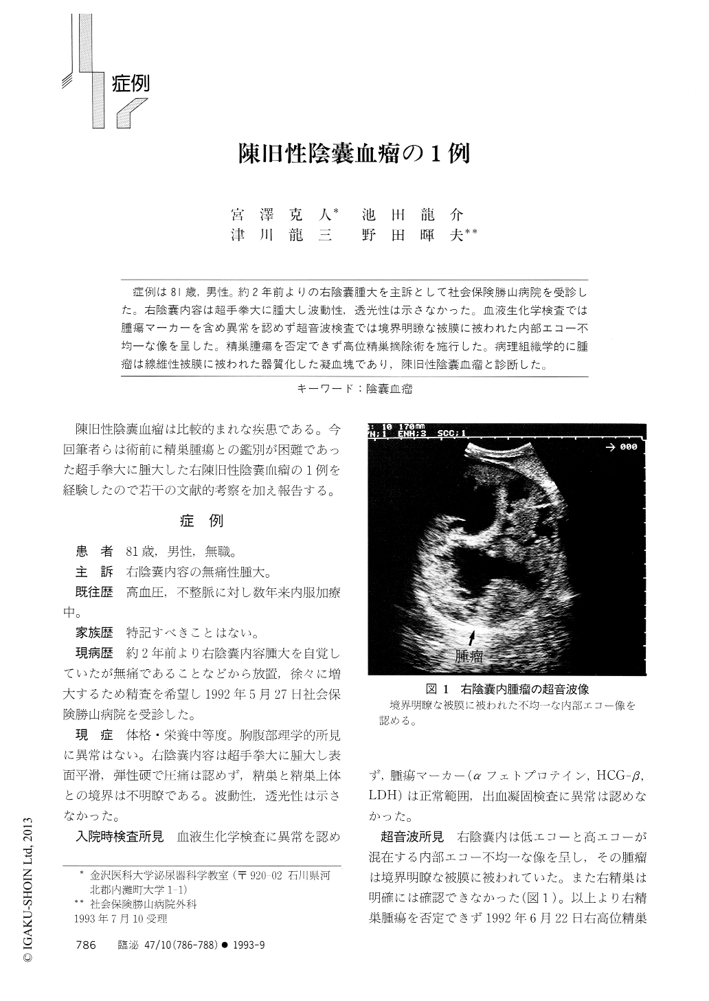
症例は81歳,男性。約2年前よりの右陰嚢腫大を主訴として社会保険勝山病院を受診した。右陰嚢内容は超手拳大に腫大し波動性,透光性は示さなかった。血液生化学検査では腫瘍マーカーを含め異常を認めず超音波検査では境界明瞭な被膜に被われた内部エコー不均一な像を呈した。精巣腫瘍を否定できず高位精巣摘除術を施行した。病理組織学的に腫瘤は線維性被膜に被われた器質化した凝血塊であり,陳旧性陰嚢血瘤と診断した。
A 81-year-old male first noticed a painless swelling in his right scrotum about 2 years before visiting our hospital. The scrotum didn't transmit light. Ultrasonography revealed an echogenic mass with an echolucent lesion in scrotum. Since it was impossible to exclude the testicular tumor completely, we performed a right high orchiectomy. The extracted mass measured 14 x 12 x 11 cm, weighed 900 g and contained old brownish black blood. Histopathological examination revealed normal testis and hematocele encapsulated with tunica vaginalis.
Copyright © 1993, Igaku-Shoin Ltd. All rights reserved.