Japanese
English
- 有料閲覧
PPV購入案内
1,320円 (1,200円+税10%) こちらは、671ページから674ページの文献です。購入には医書.jp本会員登録が必要です。
会員登録完了後に改めて上記「購入サイトへ」を選択してください。
または「購入サイトへ」選択後に「購入ログイン」、「新規会員登録」の順に進んでください。
会員登録について詳しくはをご確認ください。
- Abstract 文献概要
- 1ページ目 Look Inside
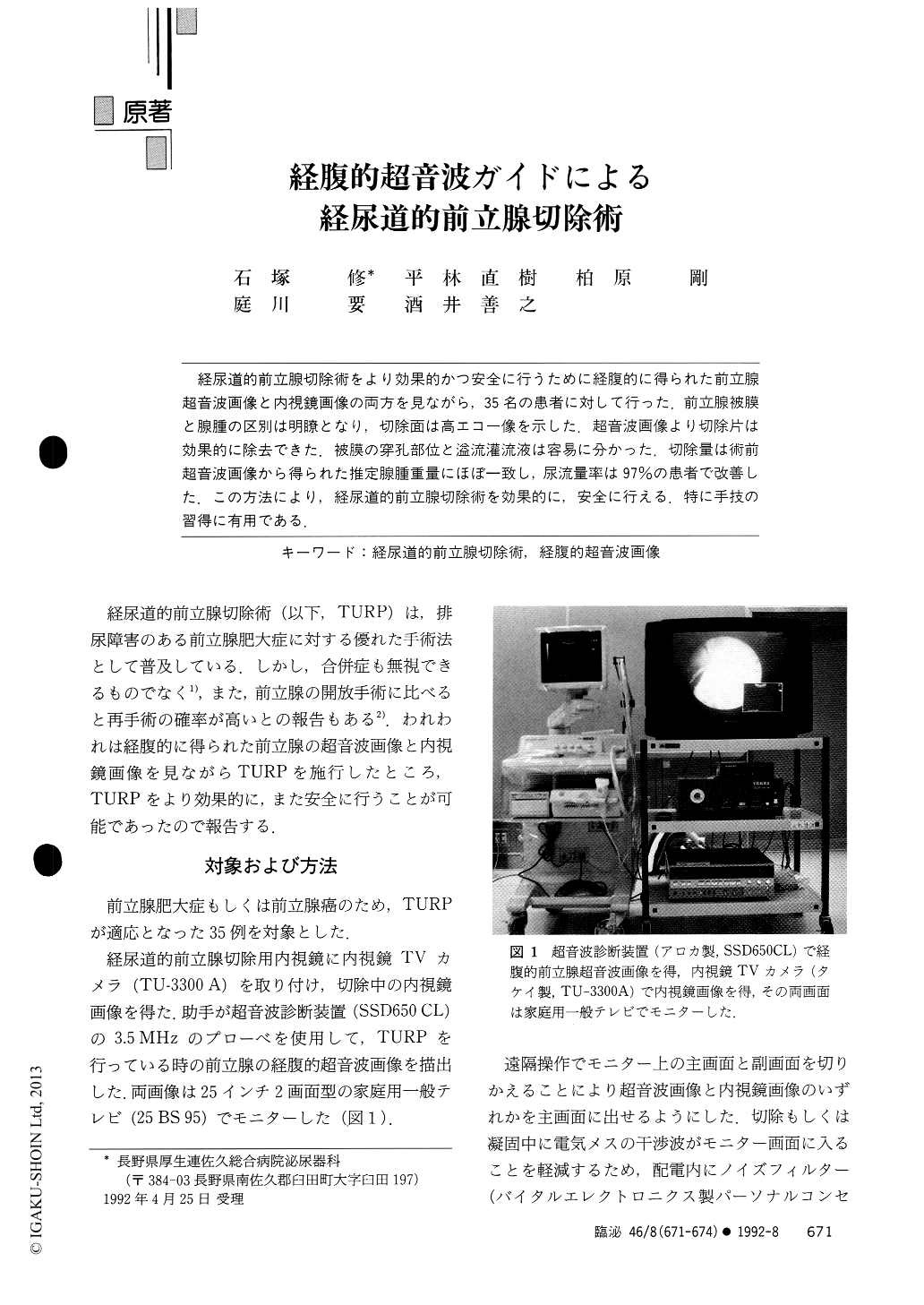
経尿道的前立腺切除術をより効果的かつ安全に行うために経腹的に得られた前立腺超音波画像と内視鏡画像の両方を見ながら,35名の患者に対して行った.前立腺被膜と腺腫の区別は明瞭となり,切除面は高エコー像を示した.超音波画像より切除片は効果的に除去できた.被膜の穿孔部位と溢流灌流液は容易に分かった.切除量は術前超音波画像から得られた推定腺腫重量にほぼ一致し,尿流量率は97%の患者で改善した.この方法により,経尿道的前立腺切除術を効果的に,安全に行える.特に手技の習得に有用である.
Observing both television monitors of endoscopy and transabdominal ultrasonographic images, weperformed transurethral resection of prostate in 35 patients. The prostatic capsule was clearly distinguishedfrom adenoma. The resection plane produced a hyperechoic image. Easy positioning of the sheath to resectedchips enabled us to evacuate them effectively. A perforated capsule and extravasated fluid were clearlyvisualized. The weight of the resected specimens was almost equal to that estimated by preoperativeultrasonography. Postoperative uroflowmetry showed an improved flow rate in 97% of patients.
Copyright © 1992, Igaku-Shoin Ltd. All rights reserved.