Japanese
English
症例
腎異物結石の1例
FOREIGN BODY STONE IN THE KIDNEY: REPORT OF A CASE
石川 英二
1
,
村田 裕
1
,
西 俊昌
1
,
添田 朝樹
1
,
松尾 光雄
1
Eiji Ishikawa
1
,
Yutaka Murata
1
,
Toshimasa Nishi
1
,
Asaki Soeda
1
,
Mitsuo Matsuo
1
1神戸市立中央市民病院泌尿器科
1Department of Urology, Kobe City General Hospital
キーワード:
腎結石
,
異物
Keyword:
腎結石
,
異物
pp.141-143
発行日 1988年2月20日
Published Date 1988/2/20
DOI https://doi.org/10.11477/mf.1413204674
- 有料閲覧
PPV購入案内
1,320円 (1,200円+税10%) こちらは、141ページから143ページの文献です。
PPV(ペイ・パー・ビュー)とは、論文(記事)単位で購入・閲覧ができるサービスです。
購入には医書.jp本会員登録が必要です。
会員登録完了後に改めて上記「購入サイトへ」を選択してください。
または「購入サイトへ」選択後に「購入ログイン」、「新規会員登録」の順に進んでください。
会員登録について詳しくはをご確認ください。
購入には医書.jp本会員登録が必要です。
会員登録完了後に改めて上記「購入サイトへ」を選択してください。
または「購入サイトへ」選択後に「購入ログイン」、「新規会員登録」の順に進んでください。
会員登録について詳しくはをご確認ください。
- Abstract 文献概要
- 1ページ目 Look Inside
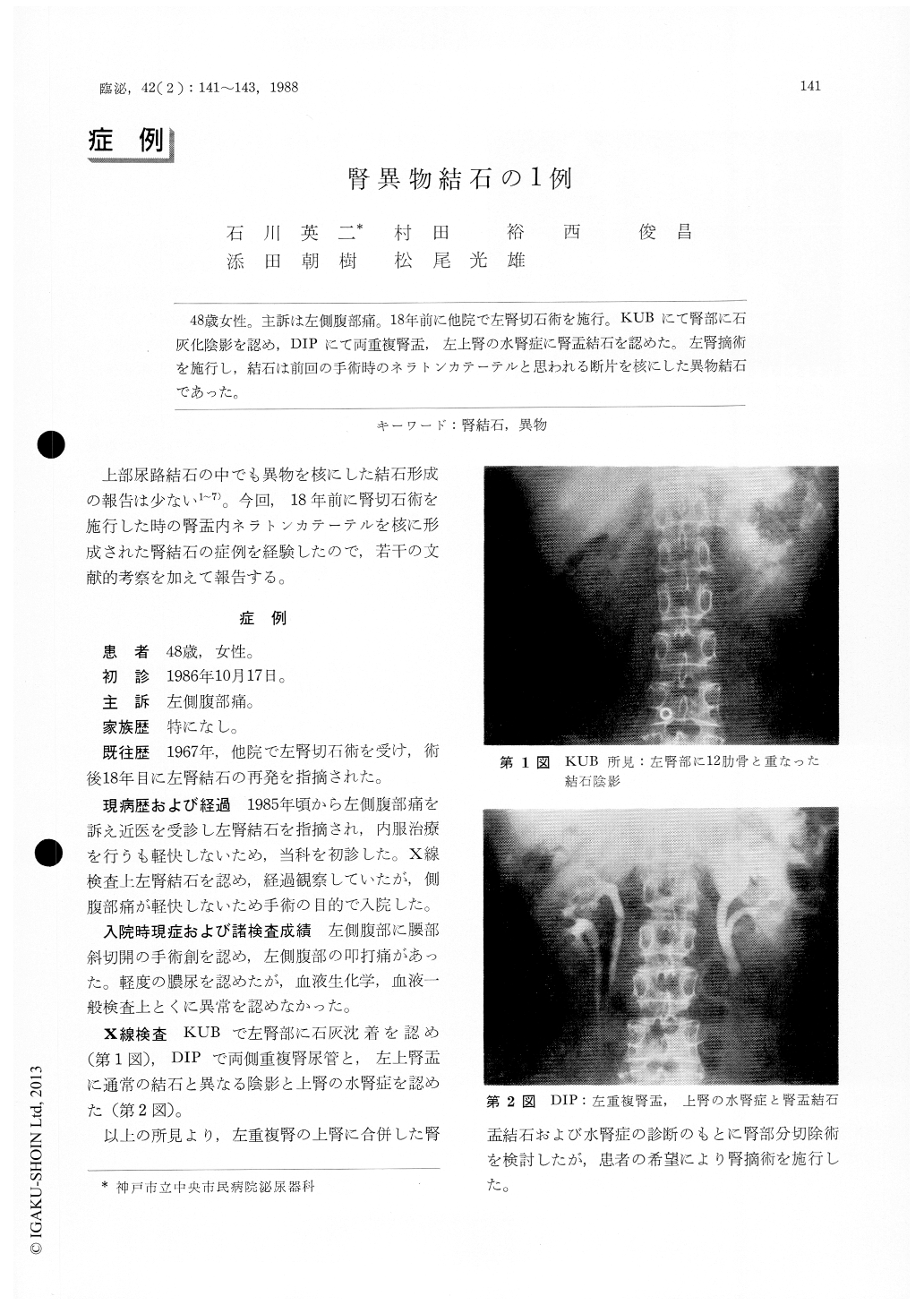
48歳女性。主訴は左側腹部痛。18年前に他院で左腎切石術を施行。KUBにて腎部に石灰化陰影を認め,DIPにて両重複腎盂,左上腎の水腎症に腎盂結石を認めた。左腎摘術を施行し,結石は前回の手術時のネラトンカテーテルと思われる断片を核にした異物結石であった。
A 48-year-old woman was admitted to our hospital with the chief complaint of left flank pain. She had left nephrolithotomy at another hospital 20 years ago. Plain X-ray film showed a calcified shadow in the upper area of the left kidney. Extretory urograms showed bilateral double pelvis and hydro-nephrosis of the upper segment of the left kidney. Left nephrectomy was performed and was disclosed foreign body stone with nuclei of the tip of rubber catheter presumably left in the previous surgery.
Copyright © 1988, Igaku-Shoin Ltd. All rights reserved.