Japanese
English
- 有料閲覧
PPV購入案内
1,320円 (1,200円+税10%) こちらは、1557ページから1561ページの文献です。購入には医書.jp本会員登録が必要です。
会員登録完了後に改めて上記「購入サイトへ」を選択してください。
または「購入サイトへ」選択後に「購入ログイン」、「新規会員登録」の順に進んでください。
会員登録について詳しくはをご確認ください。
- Abstract 文献概要
- 1ページ目 Look Inside
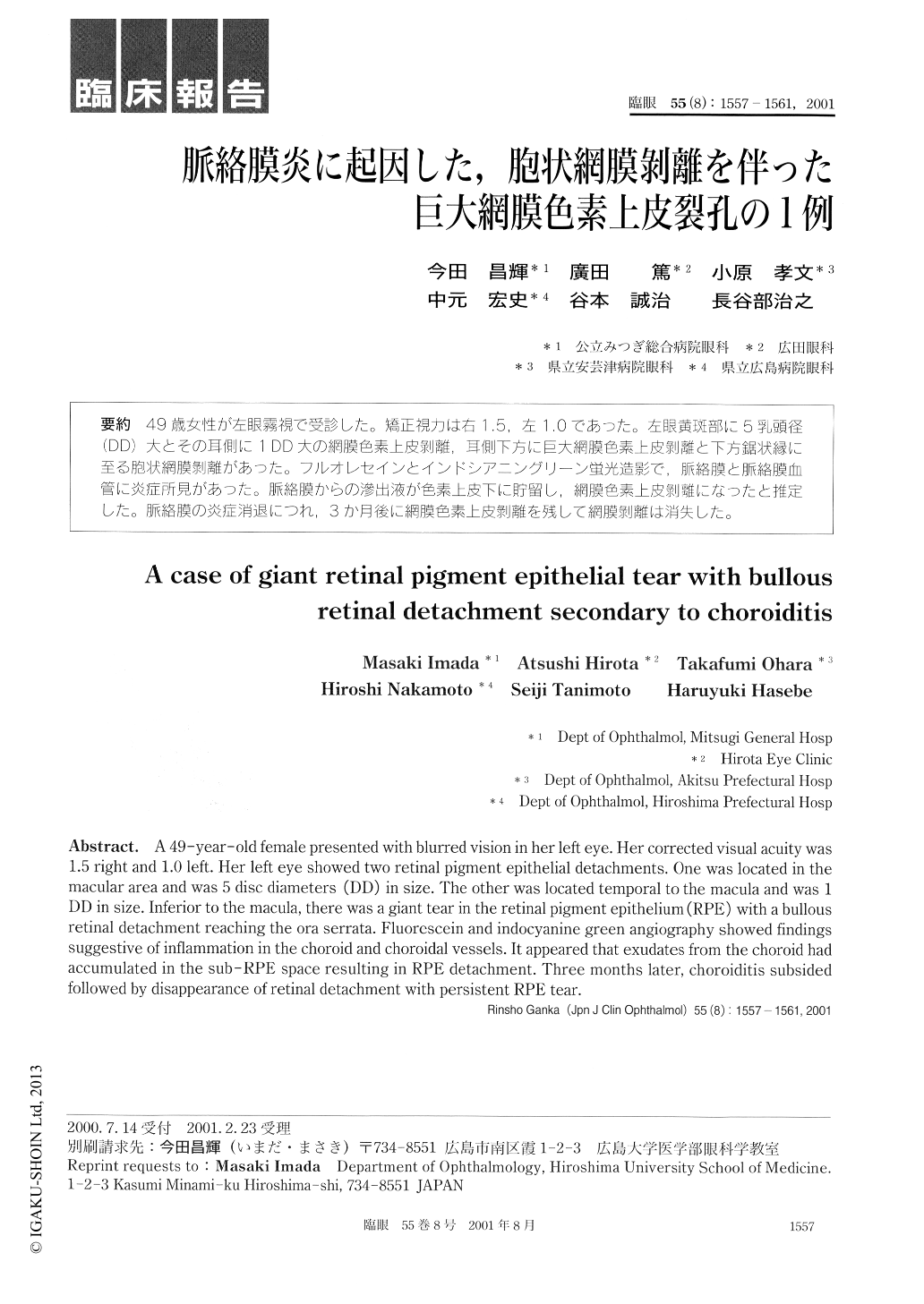
49歳女性が左眼霧視で受診した。矯正視力は右1.5,左1.0であった。左眼黄斑部に5乳頭径(DD)大とその耳側に1DD大の網膜色素上皮剥離,耳側下方に巨大網膜色素上皮剥離と下方鋸状縁に至る胞状網膜剥離があった。フルオレセインとインドシアニングリーン蛍光造影で,脈絡膜と脈絡膜血管に炎症所見があった。脈絡膜からの滲出液が色素上皮下に貯留し,網膜色素上皮剥離になったと推定した。脈絡膜の炎症消退につれ,3か月後に網膜色素上皮剥離を残して網膜剥離は消失した。
A 49-year-old female presented with blurred vision in her left eye. Her corrected visual acuity was1.5 right and 1.0 left. Her left eye showed two retinal pigment epithelial detachments. One was located in themacular area and was 5 disc diameters (DD) in size. The other was located temporal to the macula and was 1DD in size. Inferior to the macula, there was a giant tear in the retinal pigment epithelium (RPE) with a bullousretinal detachment reaching the ora serrata. Fluorescein and indocyanine green angiography showed findingssuggestive of inflammation in the choroid and choroidal vessels. It appeared that exudates from the choroid hadaccumulated in the sub-RPE space resulting in RPE detachment. Three months later, choroiditis subsidedfollowed by disappearance of retinal detachment with persistent RPE tear.
Copyright © 2001, Igaku-Shoin Ltd. All rights reserved.