Japanese
English
- 有料閲覧
PPV購入案内
1,320円 (1,200円+税10%) こちらは、1811ページから1814ページの文献です。購入には医書.jp本会員登録が必要です。
会員登録完了後に改めて上記「購入サイトへ」を選択してください。
または「購入サイトへ」選択後に「購入ログイン」、「新規会員登録」の順に進んでください。
会員登録について詳しくはをご確認ください。
- Abstract 文献概要
- 1ページ目 Look Inside
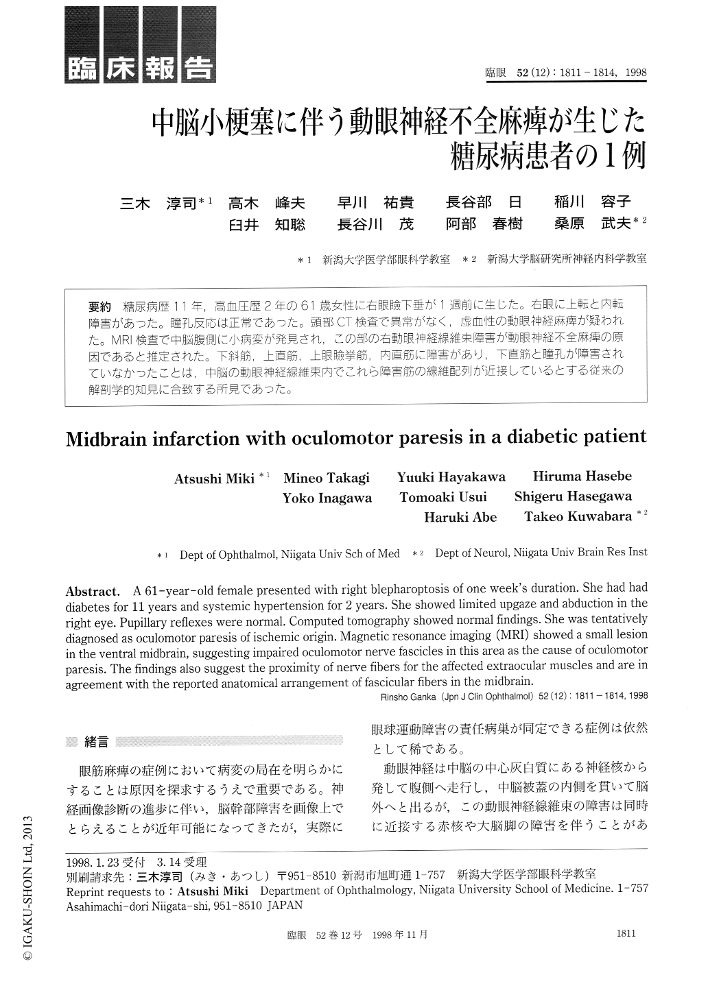
糖尿病歴11年,高血圧歴2年の61歳女性に右眼瞼下垂が1週前に生じた。右眼に上転と内転障害があった。瞳孔反応は正常であった。頭部CT検査で異常がなく,虚血性の動眼神経麻痺が疑われた。MRI検査で中脳腹側に小病変が発見され,この部の右動眼神経線維束障害が動眼神経不全麻痺の原因であると推定された。下斜筋,上直筋,上眼瞼挙筋,内直筋に障害があり,下直筋と瞳孔が障害されていなかったことは,中脳の動眼神経線維東内でこれら障害筋の線維配列が近接しているとする従来の解剖学的知見に合致する所見であった。
A 61-year-old female presented with right blepharoptosis of one week's duration. She had had diabetes for 11 years and systemic hypertension for 2 years. She showed limited upgaze and abduction in the right eye. Pupillary reflexes were normal. Computed tomography showed normal findings. She was tentatively diagnosed as oculomotor paresis of ischemic origin. Magnetic resonance imaging (MRI) showed a small lesion in the ventral midbrain, suggesting impaired oculomotor nerve fascicles in this area as the cause of oculomotor paresis. The findings also suggest the proximity of nerve fibers for the affected extraocular muscles and are in agreement with the reported anatomical arrangement of fascicular fibers in the midbrain.
Copyright © 1998, Igaku-Shoin Ltd. All rights reserved.