Japanese
English
- 有料閲覧
PPV購入案内
1,320円 (1,200円+税10%) こちらは、507ページから510ページの文献です。購入には医書.jp本会員登録が必要です。
会員登録完了後に改めて上記「購入サイトへ」を選択してください。
または「購入サイトへ」選択後に「購入ログイン」、「新規会員登録」の順に進んでください。
会員登録について詳しくはをご確認ください。
- Abstract 文献概要
- 1ページ目 Look Inside
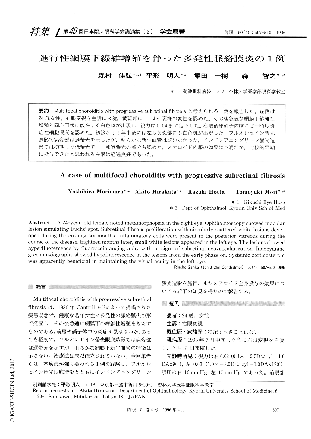
Muitifocai choroiditis with progressive subretinal fibrosisと考えられる1例を報告した。症例は24歳女性。右眼変視を主訴に来院,黄斑部にFuchs斑様の変性を認めた。その後急速な網膜下線維性増殖と同心円状に散在する白色斑が出現し,視力は0.04まで低下した。右眼後部硝子体腔には一時期炎症性細胞浸潤を認めた。初診から1年半後には左眼黄斑部にも白色斑が出現した。フルオレセイン螢光造影で病変部は過螢光を示したが,明らかな新生血管は認めなかった。インドシアニングリーン螢光造影では初期より低螢光で,一部過螢光の部分も認めた。ステロイド内服の効果は不明だが,比較的早期に投与できたと思われる左眼は経過良好であった。
A 24-year-old female noted metamorphopsia in the right eye. Ophthalmoscopy showed macular lesion simulating Fuchs' spot. Subretinal fibrous proliferation with circularly scattered white lesions devel-oped during the ensuing six months. Inflammatory cells were present in the posterior vitreous during the course of the disease. Eighteen months later, small white lesions appeared in the left eye. The lesions showed hyperfluorescence by fluorescein angiography without signs of subretinal neovascularization. Indocyanine green angiography showed hypofluorescence in the lesions from the early phase on. Systemic corticosteroid was apparently beneficial in maintaining the visual acuity in the left eye.
Copyright © 1996, Igaku-Shoin Ltd. All rights reserved.