Japanese
English
- 有料閲覧
PPV購入案内
1,320円 (1,200円+税10%) こちらは、749ページから751ページの文献です。購入には医書.jp本会員登録が必要です。
会員登録完了後に改めて上記「購入サイトへ」を選択してください。
または「購入サイトへ」選択後に「購入ログイン」、「新規会員登録」の順に進んでください。
会員登録について詳しくはをご確認ください。
- Abstract 文献概要
- 1ページ目 Look Inside
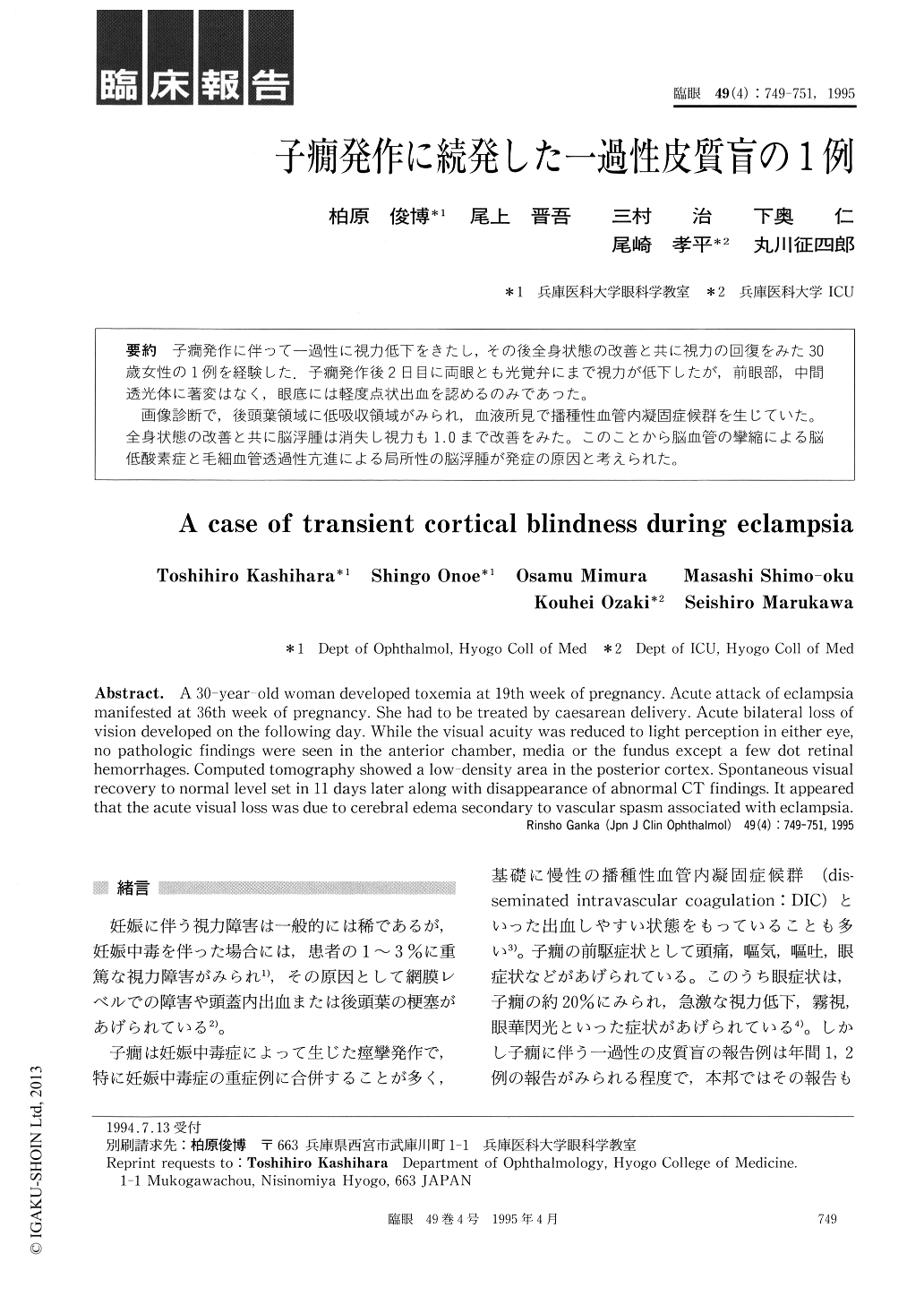
子癇痛発作に伴って一過性に視力低下をきたし,その後全身状態の改善と共に視力の回復をみた30歳女性の1例を経験した.子癇発作後2日目に両眼とも光覚弁にまで視力が低下したが,前眼部,中間透光体に著変はなく,眼底には軽度点状出血を認めるのみであった。
画像診断で,後頭葉領域に低吸収領域がみられ,血液所見で播種性血管内凝固症候群を生じていた。全身状態の改善と共に脳浮腫は消失し視力も1.0まで改善をみた。このことから脳血管の攣縮による脳低酸素症と毛細血管透過性充進による局所性の脳浮腫が発症の原因と考えられた。
A 30-year-old woman developed toxemia at 19th week of pregnancy. Acute attack of eclampsia manifested at 36th week of pregnancy. She had to be treated by caesarean delivery. Acute bilateral loss of vision developed on the following day. While the visual acuity was reduced to light perception in either eye, no pathologic findings were seen in the anterior chamber, media or the fundus except a few dot retinal hemorrhages. Computed tomography showed a low-density area in the posterior cortex. Spontaneous visual recovery to normal level set in 11 days later along with disappearance of abnormal CT findings. It appeared that the acute visual loss was due to cerebral edema secondary to vascular spasm associated with eclampsia.
Copyright © 1995, Igaku-Shoin Ltd. All rights reserved.