Japanese
English
- 有料閲覧
PPV購入案内
1,320円 (1,200円+税10%) こちらは、1157ページから1159ページの文献です。購入には医書.jp本会員登録が必要です。
会員登録完了後に改めて上記「購入サイトへ」を選択してください。
または「購入サイトへ」選択後に「購入ログイン」、「新規会員登録」の順に進んでください。
会員登録について詳しくはをご確認ください。
- Abstract 文献概要
- 1ページ目 Look Inside
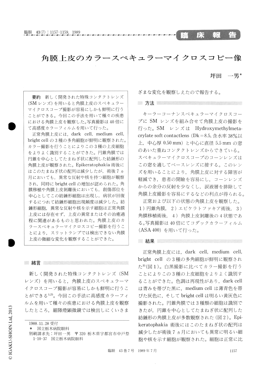
新しく開発された特殊コンタクトレンズ(SMレンズ)を用いると角膜上皮のスペキュラーマイクロスコープ撮影が容易にしかも鮮明に行うことができる。今回この手法を用いて種々の疾患における角膜上皮を観察した。写真撮影は40倍にて高感度カラーフィルムを用いて行った。
正常角膜上皮には,dark cell, medium cell, bright cellの3種の多角細胞が鮮明に観察された。カラー撮影を行うことによりこの3種の上皮細胞をよりよく識別することができた。円錐角膜では円錐を中心としてたまねぎ状に配列した紡錘形の角膜上皮が観察された。Epikeratophakia術後にはこのたまねぎ状の配列は減少したが,術後7ヵ月においても,異常な反射や核を持つ細胞が観察され,同時にbright cellの増加が認められた。角膜移植や角膜上皮剥離後においても,創傷部位を中心としてこの紡錘形細胞は出現し,病状が回復するにつれて紡錘形細胞出現頻度は減少した。紡錘形細胞,異常な反射や核を示す細胞は正常角膜上皮には存在せず,上皮の異常またはその治癒過程に関連があるものと思われた。角膜上皮のカラースペキュラーマイクロスコピー撮影を行うことにより,スリットランプでは検出できない角膜上皮の微細な変化を観察することができた。
A newly developed contact lens, SM lens, facilitates photographic documentation of the corneal epith-elium of healthy and diseased patients. I applied this method to eyes with keratoconus, following epikeratophakia or keratoplasty and with corneal erosion. Photographic documentation was made under magnification of 40-fold on reversal color film (ASA 400).
It was possible to differentiate 3 types of polygonal cells in normal corneal epithelium:dark, medium and bright cells. Diseased corneas were characterized by the presence of spindle-shaped cells and cells with abnormal reflex or nucleus. These cells seemed to be associated either with the pathological epithelium itself or with the process of recovery.
Copyright © 1989, Igaku-Shoin Ltd. All rights reserved.