Japanese
English
- 有料閲覧
PPV購入案内
1,320円 (1,200円+税10%) こちらは、861ページから867ページの文献です。購入には医書.jp本会員登録が必要です。
会員登録完了後に改めて上記「購入サイトへ」を選択してください。
または「購入サイトへ」選択後に「購入ログイン」、「新規会員登録」の順に進んでください。
会員登録について詳しくはをご確認ください。
- Abstract 文献概要
- 1ページ目 Look Inside
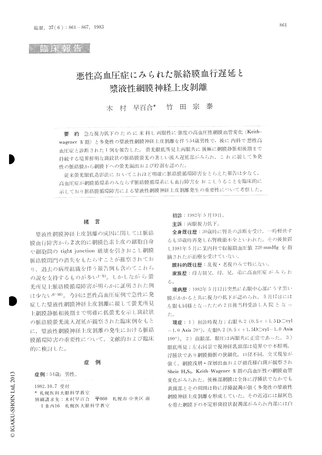
急な視力低下のために来科し両眼性に強渡の高血圧性網膜血管変化(Keith—Wagener IV期)と多発性の漿液性網膜神経上皮剥離を伴う54歳男性で,後に内科で悪性高血圧症と診断された1例を報告した。螢光眼底所見上両眼共に後極に網膜静脈相後期まで持続する境界鮮明な斑紋状の脈絡膜螢光の著しい流入遅延部がみられ,これに接して多発性の脈絡膜から網膜下への螢光漏出および貯溜を認めた。
従来螢光眼底造影法においてこれほど明確に脈絡膜循環障害をとらえた報告は少なく,高血圧症が網膜循環系のみならず脈絡膜循環系にも血行障害をおこしうることを臨床的に示しており脈絡膜循環障害による漿液性網膜神経上皮剥離発生の重要性について考察した。
A 54-year-old male visited us due to acute visual disturbance since a few days before. He had suffered from nephritis at the age of 30. Renal hypofunction and malignant hypertension seem to have been present in the intervening period.
Funduscopy revealed bilateral hypertensive vas-cular changes (4th stage by Keith-Wagener) and multiple serous retinal neuroepithelial detachments.Fluorescein fundus angiography showed multiple hypofluorescent areas arranged in mosaic pattern indicating delayed choroidal circulation. Leakage of fluorescein into the subretinal space occurred in areas neighboring the hypofluorescent ones.
Copyright © 1983, Igaku-Shoin Ltd. All rights reserved.