Japanese
English
- 有料閲覧
PPV購入案内
1,320円 (1,200円+税10%) こちらは、1487ページから1491ページの文献です。購入には医書.jp本会員登録が必要です。
会員登録完了後に改めて上記「購入サイトへ」を選択してください。
または「購入サイトへ」選択後に「購入ログイン」、「新規会員登録」の順に進んでください。
会員登録について詳しくはをご確認ください。
- Abstract 文献概要
- 1ページ目 Look Inside
緒 言
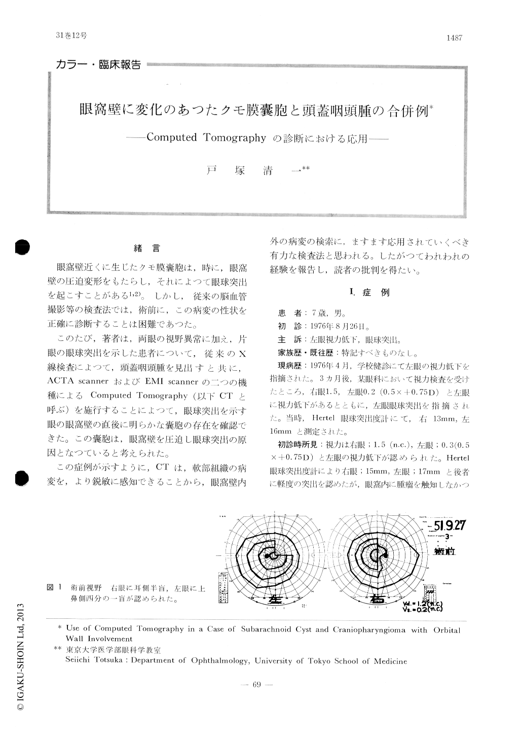
眼窩壁近くに生じたクモ膜嚢胞は,時に,眼窩壁の圧迫変形をもたらし,それによつて眼球突出を起こすことがある1,2)。しかし,従来の脳血管撮影等の検査法では,術前に,この病変の性状を正確に診断することは困難であつた。
このたび,著者は,両眼の視野異常に加え,片眼の眼球突出を示した患者について,従来のX線検査によつて,頭蓋咽頭腫を見出すと共に,ACTA scannerおよびEMI scannerの二つの機種によるComputed Tomography (以下CTと呼ぶ)を施行することによつて,眼球突出を示す眼の眼窩壁の直後に明らかな嚢胞の存在を確認できた。この嚢胞は,眼窩壁を圧迫し眼球突出の原因となつていると考えられた。
This paper describes a 7-year-old male child with decreased visual acuity, mild exophthalmos in the left eye and right homonymous hemiano-psia.
Conventional x-ray examinations revealed the presence of craniopharyngioma. Computed tomo-graphies (ACTA & EMI) showed a subarachnoid cyst in the left middle fossa and forward displ-acement of the orbital wall. The exophthalmos seemed to be due to the deformity of the orbital wall induced by the cyst. The diagnosis of the present complicated case was only possible with the use of computed tomography.
Copyright © 1977, Igaku-Shoin Ltd. All rights reserved.