Japanese
English
- 有料閲覧
PPV購入案内
1,320円 (1,200円+税10%) こちらは、587ページから591ページの文献です。購入には医書.jp本会員登録が必要です。
会員登録完了後に改めて上記「購入サイトへ」を選択してください。
または「購入サイトへ」選択後に「購入ログイン」、「新規会員登録」の順に進んでください。
会員登録について詳しくはをご確認ください。
- Abstract 文献概要
- 1ページ目 Look Inside
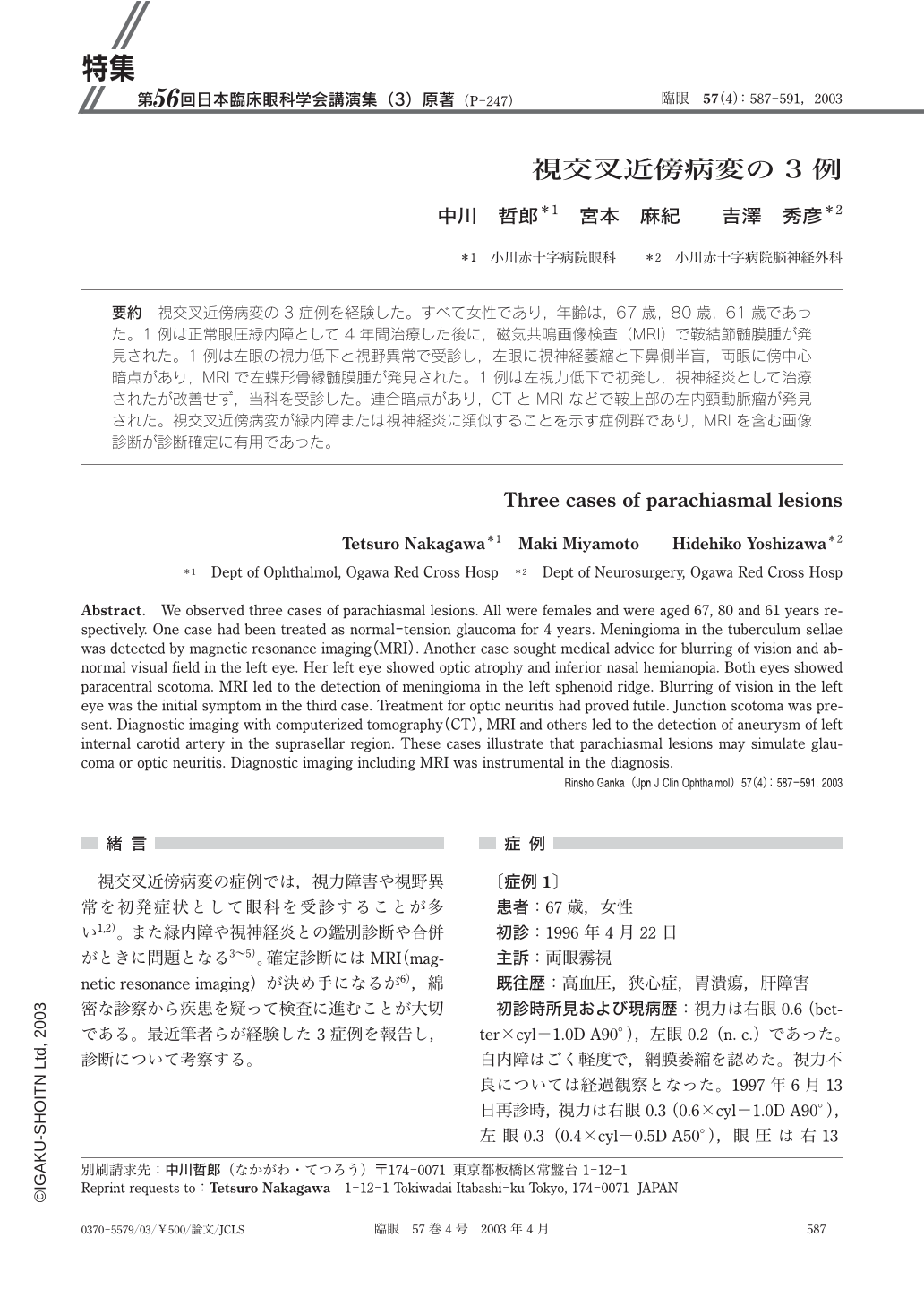
要約 視交叉近傍病変の3症例を経験した。すべて女性であり,年齢は,67歳,80歳,61歳であった。1例は正常眼圧緑内障として4年間治療した後に,磁気共鳴画像検査(MRI)で鞍結節髄膜腫が発見された。1例は左眼の視力低下と視野異常で受診し,左眼に視神経萎縮と下鼻側半盲,両眼に傍中心暗点があり,MRIで左蝶形骨縁髄膜腫が発見された。1例は左視力低下で初発し,視神経炎として治療されたが改善せず,当科を受診した。連合暗点があり,CTとMRIなどで鞍上部の左内頸動脈瘤が発見された。視交叉近傍病変が緑内障または視神経炎に類似することを示す症例群であり,MRIを含む画像診断が診断確定に有用であった。
Abstract. We observed three cases of parachiasmal lesions. All were females and were aged 67,80 and 61 years respectively. One case had been treated as normal-tension glaucoma for 4 years. Meningioma in the tuberculum sellae was detected by magnetic resonance imaging(MRI). Another case sought medical advice for blurring of vision and abnormal visual field in the left eye. Her left eye showed optic atrophy and inferior nasal hemianopia. Both eyes showed paracentral scotoma. MRI led to the detection of meningioma in the left sphenoid ridge. Blurring of vision in the left eye was the initial symptom in the third case. Treatment for optic neuritis had proved futile. Junction scotoma was present. Diagnostic imaging with computerized tomography(CT),MRI and others led to the detection of aneurysm of left internal carotid artery in the suprasellar region. These cases illustrate that parachiasmal lesions may simulate glaucoma or optic neuritis. Diagnostic imaging including MRI was instrumental in the diagnosis.
Copyright © 2003, Igaku-Shoin Ltd. All rights reserved.