Japanese
English
- 有料閲覧
PPV購入案内
1,320円 (1,200円+税10%) こちらは、479ページから482ページの文献です。購入には医書.jp本会員登録が必要です。
会員登録完了後に改めて上記「購入サイトへ」を選択してください。
または「購入サイトへ」選択後に「購入ログイン」、「新規会員登録」の順に進んでください。
会員登録について詳しくはをご確認ください。
- Abstract 文献概要
- 1ページ目 Look Inside
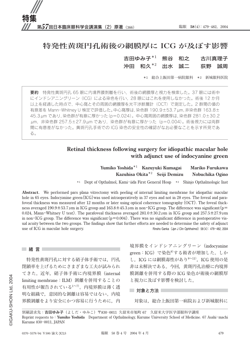
特発性黄斑円孔65眼に内境界膜剝離を行い,術後の網膜厚と視力を検索した。37眼には術中にインドシアニングリーン(ICG)による染色を行い,28眼にはこれを使用しなかった。術後12か月以上を経過した時点で,中心窩とその周囲の網膜厚を光干渉断層計(OCT)で測定した。2群間の値の有意差をMann-Whitney U検定で評価した。中心窩厚は,染色群190.9±53.7μm,非染色群163.8±45.3μmであり,染色群が有意に厚かった(p=0.024)。中心窩周囲の網膜厚は,染色群281.0±30.2μm,非染色群257.5±27.9μmであり,染色群が有意に厚かった(p=0.004)。術後視力には両群間に有意差がなかった。黄斑円孔手術でのICG染色の安全性の確認がなお必要なことを示す所見である。
We performed pars plana vitrectomy with peeling of internal limiting membrane for idiopathic macular hole in 65 eyes. Indocyanine green(ICG)was used intraoperatively in 37 eyes and not in 28 eyes. The foveal and parafoveal thickness was measured after 12months or later using optical coherence tomography(OCT). The foveal thickness averaged 190.9±53.7μm in ICG group and 163.8±45.3μm in non-ICG group. The difference was significant(p=0.024,Mann-Whitney U test). The parafoveal thickness averaged 281.0±30.2μm in ICG group and 257.5±27.9μm in non-ICG group. The difference was significant(p=0.004). There was no significant difference in postoperative visual acuity between the two groups. The findings show that further efforts are needed to determine the safety of adjunct use of ICG in macular hole surgery.
Copyright © 2004, Igaku-Shoin Ltd. All rights reserved.