Japanese
English
- 有料閲覧
PPV購入案内
1,320円 (1,200円+税10%) こちらは、1401ページから1406ページの文献です。購入には医書.jp本会員登録が必要です。
会員登録完了後に改めて上記「購入サイトへ」を選択してください。
または「購入サイトへ」選択後に「購入ログイン」、「新規会員登録」の順に進んでください。
会員登録について詳しくはをご確認ください。
- Abstract 文献概要
- 1ページ目 Look Inside
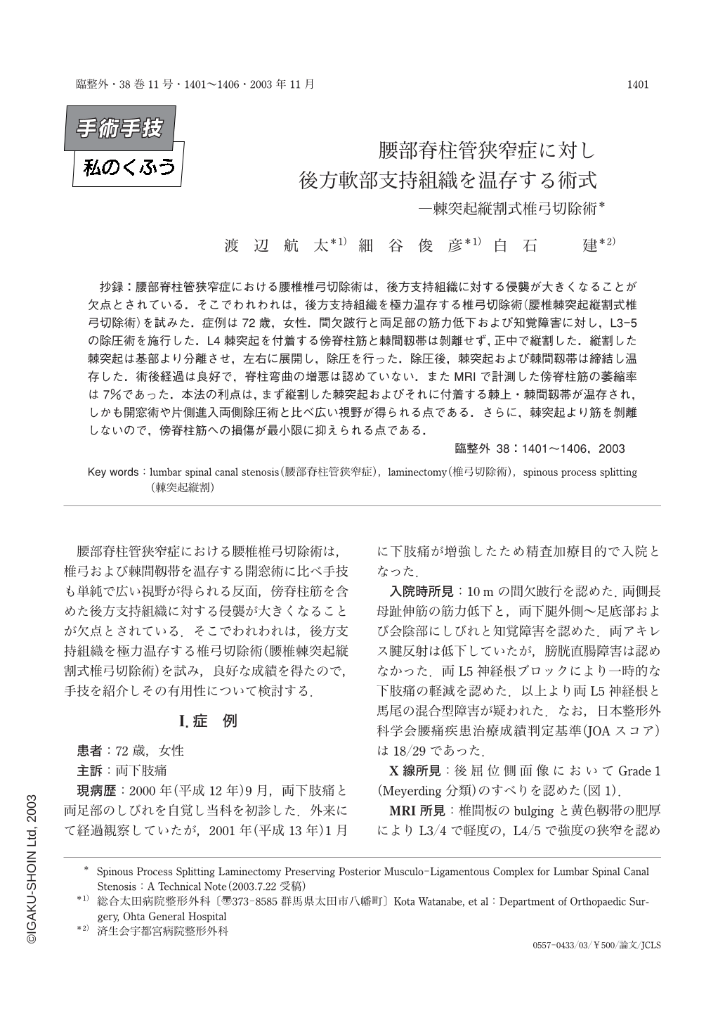
抄録:腰部脊柱管狭窄症における腰椎椎弓切除術は,後方支持組織に対する侵襲が大きくなることが欠点とされている.そこでわれわれは,後方支持組織を極力温存する椎弓切除術(腰椎棘突起縦割式椎弓切除術)を試みた.症例は72歳,女性.間欠跛行と両足部の筋力低下および知覚障害に対し,L3-5の除圧術を施行した.L4棘突起を付着する傍脊柱筋と棘間靱帯は剥離せず,正中で縦割した.縦割した棘突起は基部より分離させ,左右に展開し,除圧を行った.除圧後,棘突起および棘間靱帯は締結し温存した.術後経過は良好で,脊柱弯曲の増悪は認めていない.またMRIで計測した傍脊柱筋の萎縮率は7%であった.本法の利点は,まず縦割した棘突起およびそれに付着する棘上・棘間靱帯が温存され,しかも開窓術や片側進入両側除圧術と比べ広い視野が得られる点である.さらに,棘突起より筋を剝離しないので,傍脊柱筋への損傷が最小限に抑えられる点である.
In conventional laminectomy of the lumbar spine, the paravertebral muscles are invariably detached from the posterior arches of the lumbar vertebrae. Furthermore, the posterior midline ligaments as well as the dorsolumbar fascia lose their anchoring points as a result of removal of the spinous processes. Such intraoperative injuries to the posterior extensor mechanism of the lumbar spine often lead to spinal malalignment or instability associated with weakness of the trunk muscles. To minimize the injuries, we developed a new lumbar laminectomy procedure adopting Shiraishi's less invasive technique which was originally designed for cervical spinal canal decompression. In the new procedure for the lumbar spine, the laminae are exposed by longitudinally splitting each spinous process into two halves with its muscular and ligamentous attachments kept undisturbed. After separating the split spinous processes from the posterior arches, the cauda equina and the nerve roots are decompressed by laminectomy. Before closing the wound, the split halves of each spinous process are re-approximated with a strong suture. We performed this new procedure on a 72-year-old lady with severe canal stenosis between L3 and L5. Neurological recovery has been successful for 2 years postoperatively with the spinal alignment well maintained on plain radiographs. Postoperative cross-sectional area of the paravertebral muscles measured on axial MRI was preserved 93% of the preoperative one. The advantages of the procedure are 1)the midline osteoligamentous structures are preserved, 2)a large surgical workspace is obtained as compared to fenestration, and 3)preservation of the muscular attachments to the spinous processes minimizes damage to the paravertebral muscles.
Copyright © 2003, Igaku-Shoin Ltd. All rights reserved.