Japanese
English
- 有料閲覧
PPV購入案内
1,320円 (1,200円+税10%) こちらは、363ページから367ページの文献です。購入には医書.jp本会員登録が必要です。
会員登録完了後に改めて上記「購入サイトへ」を選択してください。
または「購入サイトへ」選択後に「購入ログイン」、「新規会員登録」の順に進んでください。
会員登録について詳しくはをご確認ください。
- Abstract 文献概要
- 1ページ目 Look Inside
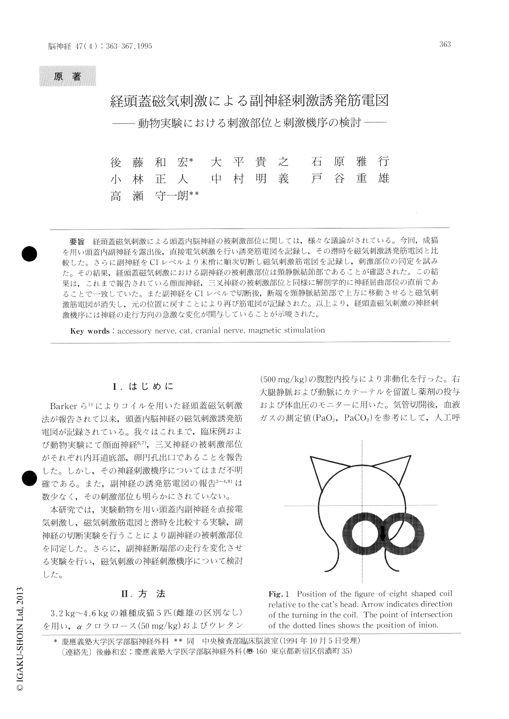
経頭蓋磁気刺激による頭蓋内脳神経の被刺激部位に関しては,様々な議論がされている。今回,成猫を用い頭蓋内副神経を露出後,直接電気刺激を行い誘発筋電図を記録し,その潜時を磁気刺激誘発筋電図と比較した。さらに副神経をC1レベルより末梢に順次切断し磁気刺激筋電図を記録し,刺激部位の同定を試みた。その結果,経頭蓋磁気刺激における副神経の被刺激部位は頸静脈結節部であることが確認された。この結果は,これまで報告されている顔面神経,三叉神経の被刺激部位と同様に解剖学的に神経屈曲部位の直前であることで一致していた。また副神経をC1レベルで切断後,断端を頸静脈結節部で上方に移動させると磁気刺激筋電図が消失し,元の位置に戻すことにより再び筋電図が記録された。以上より,経頭蓋磁気刺激の神経刺激機序には神経の走行方向の急激な変化が関与していることが示唆された。
The site where transcranial magnetic stimulation excites the accessory nerve was studied in 5 cats. Transcranial magnetic stimulation of the accessory nerve was recorded from the right trapezius. The accessory nerve was stimulated electrically at the C1 level, jugular tubercle and jugular foramen. The latencies of the compound muscle action potentials (CMAPs) for each portion were measured and compared with the magnetic response, which was coincidental with that of the jugular tubercle.
Copyright © 1995, Igaku-Shoin Ltd. All rights reserved.