Japanese
English
- 有料閲覧
PPV購入案内
1,320円 (1,200円+税10%) こちらは、289ページから294ページの文献です。購入には医書.jp本会員登録が必要です。
会員登録完了後に改めて上記「購入サイトへ」を選択してください。
または「購入サイトへ」選択後に「購入ログイン」、「新規会員登録」の順に進んでください。
会員登録について詳しくはをご確認ください。
- Abstract 文献概要
- 1ページ目 Look Inside
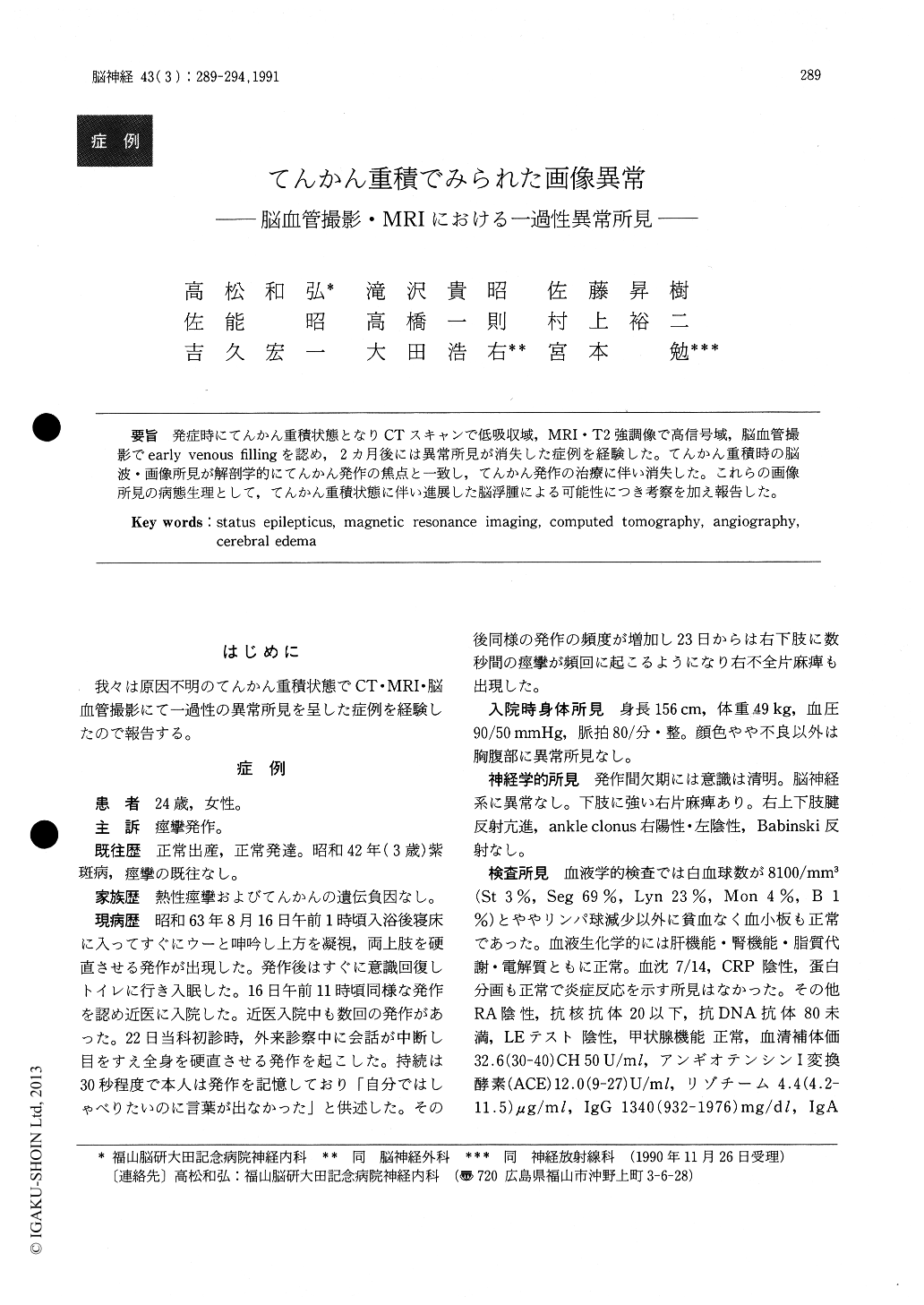
発症時にてんかん重積状態となりCTスキャンで低吸収域,MRI・T2強調像で高信号域,脳血管撮影でearly venous fillingを認め,2カ月後には異常所見が消失した症例を経験した。てんかん重積時の脳波・画像所見が解剖学的にてんかん発作の焦点と一致し,てんかん発作の治療に伴い消失した。これらの画像所見の病態生理として,てんかん重積状態に伴い進展した脳浮腫による可能性につき考察を加え報告した。
We reported transient changes in computed tomo-graphy (CT) , angiography and magnetic resonanceimaging (MRI) scans in a patient with status epile-pticus, referred to us with a tentative diagnosis ofneoplasma based on CT, MRI and angiographicfindings, MRI showed increased signal intensity, andCT showed decreased left hemisphere attenuationwithout enhancement. Two months later, resolutionof these radiological and clinical abnormalities hadbeen attained. The transient CT and MRI changesprobably represented focal cerebral edema, develop-ing during focal status epilepticus.
Copyright © 1991, Igaku-Shoin Ltd. All rights reserved.