Japanese
English
- 有料閲覧
- Abstract 文献概要
- 1ページ目 Look Inside
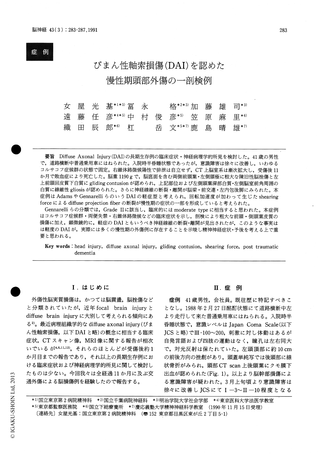
Diffuse Axonal Injury(DAI)の長期生存例の臨床症状・神経病理学的所見を検討した。41歳の男性で,道路横断中普通乗用車にはねられた。入院時半昏睡状態であったが,意識障害は徐々に改善し,いわゆるコルサコフ症候群の状態で固定。右錐体路徴候陽性で排泄は自立せず,CT上脳室系は漸次拡大し,受傷後11か月で敗血症により死亡した。脳重1190gで,脳底面を含む両側前頭葉・左側頭極に粗大な陳旧性脳挫傷と左上前頭回皮質下白質にgliding contusionが認められ,上記部位および左側頭葉深部白質・左側脳室前角周囲の白質に線維性gliosisが認められた。さらに神経線維の断裂・離開が脳梁・前交連・左内包後脚にみられた。本症例はAdamsやGennarelliらのいうDAIの軽症型と考えられ,回転加速度が加わって生じたshearingforceによるdffuse projection fiberの断裂が慢性期の症状の一部を形成していると考えられた。
Gennarelliらの分類では,Grade IIに該当し,臨床的にはmoderate typeに相当すると思われた。本症例はコルサコフ症候群・両便失禁・右錐体路徴候などの臨床症状を示し,剖検により粗大な前頭・側頭葉皮質の損傷に加え,顕微鏡的に,軽症のDAIともいうべき神経線維の断裂・離開が見出されたが,このような事実はは軽度のDAIが,実際には多くの慢性期の外傷例に存在することを示唆し精神神経症状・予後を考える上で重要と思われる。
The authors reported a clinico-pathological case survived 11 months after a traffic accident. A 41-year-old man had been hit by a motorcar and was found in a state of semicoma. On admission, his consciousness level was III-100 to 200 (Japan Coma Scale). Pupils were isocoric ; light reflex was pres-ent. Linear fracture of occipital bone was disclosed by Skull X-ray and subarachnoid hemorrhage was revealed on CT scan. This comatous state, lasting 24 hours, slowly improved and eventually he presented the so-called Korsakoff's syndrome until his death. He could not recognized his relatives, only uttered some meaningless words. He was un-able to obey simple verbal orders. The patient was incontinent and right pyramidal sign was positive. On repeated CT scans, cerebral ventricles gradually increased in size ; especially the enlargement of the fourth ventricle was remarkable. He expired of septic shock caused by bed sores.
At autopsy brain weighed 1190g. Old gloss contusional scars were observed on the bilateral frontal lobes including the orbital area and on the left temporal pole. Gliding contusions were revealed in the subcortical white matter beneath the left superior frontal convolution. Fibrillary gliosis was noted in this region, the deep white matter under-lying the left temporal pole and the tissue surround-ing the anterior horn of the left lateral ventricle.
Nerve fibers were fragmented and lacerated at corpus callosum, anterior commissure and posterior limb of the left internal capsule. Bilateral pyramidal tracts showed mild myelin pallor at the brainstem. Loss of Purkinje cells were observed.
This case would correspond to mile type of diffuse axonal injury proposed by Adams and Gennarelli. Disruption of diffuse projection fibers caused by angular acceleration producing shear strain might play a part in the formation of clinical symptoms (including dementia, in a wide sense). According to the classification of Gennarelli this case deserves pathologically the grade II and clinically the moder-ate type. Absence of axonal retraction ball is prob-ably due to long survival. It was considered that ventricular dilatation resulted from reduction of the bulk of white matter. This case represents a mild diffuse axonal injury and the number of such autopsy cases as this is very limited. Further clinico-pathological investigations of the cases like this may contribute to elucidating the mechanism and the outcome of head trauma.

Copyright © 1991, Igaku-Shoin Ltd. All rights reserved.


