Japanese
English
- 有料閲覧
PPV購入案内
1,320円 (1,200円+税10%) こちらは、739ページから746ページの文献です。購入には医書.jp本会員登録が必要です。
会員登録完了後に改めて上記「購入サイトへ」を選択してください。
または「購入サイトへ」選択後に「購入ログイン」、「新規会員登録」の順に進んでください。
会員登録について詳しくはをご確認ください。
- Abstract 文献概要
- 1ページ目 Look Inside
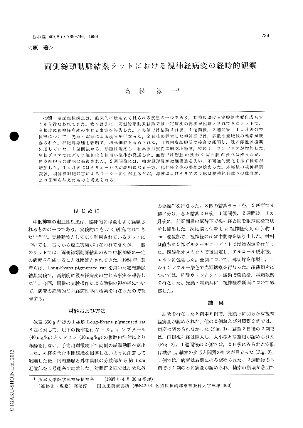
抄録 脳虚血性疾患は,臨床的に最もよく見られる疾患の一つであり,動物における実験的病変作成も古くから行なわれてきた。我々は先に,両側総頸動脈結紮では一定病変の再現が困難とされてきたラットで,高頻度に視神経病変の生じる事実を報告した。本実験では結紮2日後,1週間後,2週間後,1カ月後の視神経について,光顕・電顕による検索を行なった。2日後の腫大した視神経では,多数の空胞状の軸索が観察された。細胞外浮腫も著明で,壊死細胞も認められた。血管内皮細胞間の接合は離開し,既に浮腫は極期に達していた。1週間後から,浮腫は退潮し,軸索原形質内に細胞小器管,特にミトコンドリアが増加した。星状グリアではグリア細線維と粗面小胞体が発達した。血管では管腔の変形や周囲腔の変化は残ったが,内皮細胞間の離開は修復された。2週間後には,軸索原形質が微細構造を失い,不可逆的変化を示す軸索が増加した。1ヵ月後にはグリオーシスが著明になる一方,視神経全体の萎縮が始まった。本実験の視神経病変は,視神経細胞障害によるワーラー変性が主体だが,浮腫およびグリアの反応は視神経自体への虚血が,より影響を与えたものと考えられる。
Ischemic desease is common in the central ner-vous system, and has been extensively studied both clinically and neuropahtologically. Experi-mental models have been designed for the repro-duction of cerebral infarct. Advanced optic nerve atrophy was recently described in Long-Evans pigmented rat 16 weeks following bilateral carotid artery ligation. Marked change in the retina and its vessels had previously been observed by the fundoscopy and the fluorescein angiography. Serial alteration of the optic nerve was studied on the same experimental model for one month ; each two rats for two days, one week, two weeks, one month after the operation, in which the common carotid arteries were ligated just before the bifur-cation. A mid-portion of the optic nerve attached to the brain was dissected out and cross-sections were prepared to examine under the light and the electron microscope. Unequivocal pathology was demonstrated in six among eight rats operated. There was no significant change both in remain-ing two rats and two sham-operated controls. Two-days-postoperative rats developed obvious optic nerve swelling with vacuolar appearance.They consisted of numerous distended axons, con-taining empty space or flocculus materials. Both intra- and extracellular edema were seen at this time, when vascular beds were transiently dis-rupted. Necrotic cells and other residual materials were also present in the wide interstitial space. In the following one or two weeks, the edema disappeared and many distended axons contained a lot of organelles, such as mitochondria and/or neurofilaments. In the same period severely dama-ged axons collapsed, occasionally concomitant with homogenous dense axoplasm. There were as many protoplasmic astrocytes as phagocytic cells contain-ing remnants of the tissue or lipid droplets. Vascular beds appeared to be reconstructed. And then, the majority of the optic nerves were pro-gressively replaced with compactly packed glial processes during the latter half of the experiment, whereas still being present a variety of the alte-ration, even normal axons. Myelin sheath was preserved relatively longer, resulting in the pre-sence of axons with the empty center. The patho-logy of this experiment could be mainly due to Wallerian degeneration secondary to the damage of the ganglion cell in the retina. However di-rect ischemic effect to the optic nerve itself might accelerated the interstitial changes such as floody edema and astroglial reaction. The early optic nerve swelling was compared with the papillede-ma. Author felt this model would be as much as useful for studying ischemic disease in the central nervous system as other known ischemic models.
Copyright © 1988, Igaku-Shoin Ltd. All rights reserved.