Japanese
English
- 有料閲覧
PPV購入案内
1,320円 (1,200円+税10%) こちらは、661ページから667ページの文献です。購入には医書.jp本会員登録が必要です。
会員登録完了後に改めて上記「購入サイトへ」を選択してください。
または「購入サイトへ」選択後に「購入ログイン」、「新規会員登録」の順に進んでください。
会員登録について詳しくはをご確認ください。
- Abstract 文献概要
- 1ページ目 Look Inside
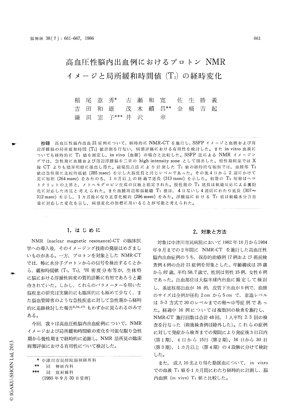
抄録 高血圧性脳内出血21症例について,経時的にNMR-CTを施行し,SSFPイメージと血腫および周辺浮腫脳の局所緩和時間(T1)値計測を行ない,病態評価における有用性を検討した。またin vitro血液についても経時的にT1値を測定し,in vivo (血腫)の場合と比較した。SSFP法によるNMRイメージングでは,急性期に血腫および周辺浮腫脳を二重のhigh intensity zoneとして描出した。慢性期病巣ではX線CTよりも境界明瞭に描出し得た。磁場焦点法により計測したT1値の経時的な観察では,血腫部T1値は急性期に比較的低値(285msec)を示し大脳皮質と同じレベルであった。その後4日から2週にかけて更に短縮(264msec)をみたのち,1カ月以上の経過で延長(313 msec)を示した。初期のT1短縮はヘマトクリットの上昇と,メトヘモグロビン生成の反映と推定された。慢性期のT1延長は組織反応による嚢胞化に対応した所見と考えられた。また血腫周辺部脳組織T1値は,4日ないし4週間にわたり延長(307〜312msec)を示し,1カ月後になり正常化傾向(296 msec)をみた。浮腫脳におけるT1値は組織水分含有量に対応した変化を示し,病態変化の指標に用いることが可能と考えられた。
The present study describes time courses in tis-sue T1 values, as well as in NMR imagings, as-sociated with hypertensive intracerebral hematoma (ICH). Non-operative 21 cases of ICH were exa-mined by FONAR QED 80-α NMR system, which possessed dual modes of image display and focal T1 measurment (static magnetic field: 433 gauss). As the first step of examination SSFP images are displayed and then, at the regions of interest, absolute values of T1 are measured by field focusing technique.
The extent of ICH was revealed as high density zone in NMR imaging, occasionally represented much wider extent of high density area than the finding on X-ray CT. Prolonged T1 values were obtained from such high density zone. This wide-spread high density area was regarded to reflect the spread of perifocal brain edema.
T1 value of the hematoma itself was rather shortened in its initial phase within 2 weeks, thereafter followed by prolongation in the time lapse. This seemed to reflect the alterations in the properties of hematoma such as clot formation in earlier phase and resolution in later phase. On the contrary, T1 in the brain tissue surrounded to hematoma was apparently prolonged in the early phase within 2 weeks, representing the max-imal values of 312 msec arround 2 to 4 weeks after the onset, and then gradually normalized in the period over 1 month. This alteration in tissue T1 likely represents the processes of edema formation and its regression in perifocal zone. T1 values measured in perifocal region might be available for the evaluation of edema state in association with cerebrovascular accident.
Copyright © 1986, Igaku-Shoin Ltd. All rights reserved.